Edited by Dr. Peg Miller, ADDL Pathologist
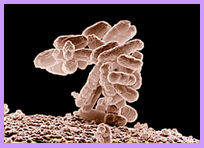 Mastitis is one of the most economically significant diseases in dairy cattle, causing decreased milk production and increased treatment, labor, and veterinary costs. Though numerous contagious and environmental mastitis pathogens exist, coliform bacteria are the most common cause of fatal mastitis in all types of production systems. Coliform mastitis pathogens, mainly Escherichia coli, Klebsiella sp., and Enterobacter sp., are lactose-fermenting, gram-negative rods in the Enterobacteriaceae family. The prevalence of coliform infection is low, constituting only 2-4% of mastitis cases; however, severe clinical mastitis is common and frequently fatal within days of bacterial invasion. The massive release of lipopolysaccharide (LPS) endotoxin causes edema, hemorrhage, and necrosis of mammary tissue along with systemic effects. With severe systemic disease, early diagnosis and immediate initiation of treatment is essential to overcome the endotoxic effects and increase cow survival rates.
 Unlike many of the common contagious mastitis pathogens, E.coli, Klebsiella, and Enterobacter are opportunistic bacteria found in feces and are ubiquitous in even the most well-managed dairy environments. Though coliforms may invade and cause mastitis through hematogenous or percutaneous routes, the most frequent route of invasion is through the teat canal. Infections typically occur in multiparous cows from two weeks before until two weeks after calving when the teat canal is most vulnerable to bacterial invasion and the cow is immunocompromised. Coliform mastitis also commonly occurs close to the drying off period if the keratin plug fails to form in the teat canal or no dry-cow treatment is pursued by the producer. Use of sawdust bedding, low somatic cell count, and damage of the teat ends by overmilking or poor milking machine maintenance, are all risk factors for coliform mastitis. Infection is most common in cows housed in total confinement or dry lots, and is rarely observed in pastured cows.
In most cows, coliform bacteria do not cause clinical mastitis upon invading the gland. The bacteria briefly multiply, but infections are cleared within one to two days by host defenses, such as lactoferrin, lysozymes, lactoperoxidase, and immunoglobulins, in addition to regular milkings. However, susceptible cows may become infected after a single exposure to the teat canal. After the initial introduction into the canal, the bacteria quickly invade the teat cistern and proliferate with release of endotoxins. The endotoxins cause an increase in vascular permeability, a marked influx of neutrophils, and extensive cytokine release resulting in necrosis and severe vascular leakage. The severity of the mastitis depends on the number of neutrophils within the milk and invading the gland, susceptibility of the bacteria to host defenses, and the degree of endotoxic injury. Unlike Staphylococcus aureus, coliforms typically do not invade the mammary parenchyma, but the decrease in defenses associated with coliform infections may lead to secondary infections with invasive pathogens.
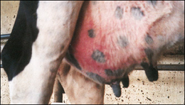
The common clinical signs of coliform mastitis are associated with the release of endotoxin and include swelling of the udder, watery milk with small flakes, decreased milk production, fever, and anorexia. Peracute cases may result in the pyrexia, tachycardia, and shock of endotoxemia. Infections typically affect only one quarter, but cause clinical mastitis in up to 90% of cases and are usually associated with systemic disease. Some cases of coliform mastitis may become chronic, but most more than 50% of infections end within ten days. Considering the typical timing and systemic effects of coliform mastitis, the differential diagnosis might include parturient hypocalcemia or carbohydrate overload with rumen acidosis. With the mammary reaction, environmental streptococcal infections, Streptococcus agalactiae, or Staphylococcus aureus may also be considered in the differential diagnosis.
A clinical diagnosis of coliform mastitis is usually based on clinical signs, culture of coliform organisms from milk, and a high somatic cell count. In many cases, culturing for coliform bacteria may be unrewarding because host defenses can clear the bacteria from the gland in one to two milkings. Streaking blood agar and MacConkey agar plates with 0.01 ml and 0.1 ml of milk, respectively, may aid in obtaining detectable growth and a definitive diagnosis. If the animal has already succumbed to the effects of endotoxemia, bacteriology of chilled mammary tissue and regional lymph nodes along with histologic evaluation of formalin-fixed mammary tissue are diagnostic in confirming coliform mastitis.
Clinical pathology findings in peracute coliform mastitis include hemoconcentration and a marked leukopenia characterized by neutropenia and a degenerate left shift. A moderate lymphopenia, monocytopenia, and thrombocytopenia may also be noted. Serum biochemistry in severe cases may indicate uremia, high aspartate aminotransferase, and a metabolic acidosis. California Mastitis Test scores of +3 and a Somatic Cell Count of 14-25,000,000 cells/ml of milk support the diagnosis.
Gross lesions of coliform mastitis consist of marked edema, hemorrhage, and necrosis of mammary tissues. Cell injury results in the sequestration of necrotic tissue surrounded by hyperemia. Secretions in the cisterns are usually serosanguineous with small clots of fibrin or coagulated casein, which may obstruct the ducts. If the cow survives the initial endotoxemia, the necrotic mammary tissue will be sequestered from the unaffected tissue and sloughed. Gangrenous mastitis is usually not caused by coliform infection alone, but secondary invasion of Staphylococcus aureus may lead to gangrenous change.
Histologically, the leukocytic infiltration of coliform mastitis is centered around interlobular ducts. Accumulation of fibrin and necrotic debris can obstruct smaller intralobular ducts. Mammary acini are filled with serous fluid, but few leukocytes are observed in the acini or in the septa. Interlobular septa are edematous; dilated lymphatic vessels contain fibrin plugs. Marked hemorrhage may be noted in the stroma and acini. In less severe cases, chronic lesions may develop in the lactiferous sinus including hyperplasia, disorganization, and changes in the epithelial lining.
Even though the bacterial invasion begins in the mammary gland, treatment of coliform mastitis must also combat the systemic endotoxemia. Broad spectrum intravenous antimicrobials are given to severely affected cows to reduce the bacteremia which may be present in up to 48% of coliform mastitis cases. Intramammary infusions of antibiotics typically do not affect the outcome of coliform mastitis cases but may be beneficial in decreasing pathogenic gram-positive organisms within the mammary gland. Stripping of the affected quarter several times a day, in conjunction with oxytocin injections, has also been utilized to reduce the number of bacteria and endotoxin in the gland. Anti-inflammatory and fluid therapy may also be necessary to prevent shock and maintain overall hydration status.
Prevention of coliform mastitis is difficult because Escherichia coli, Klebsiella, and Enterobacter are ubiquitous in even the most well-managed dairies. Culturing individual milk samples is important to identify the major pathogens in the herd. Improving pre-milking hygiene and frequent evaluation of milking machine function may reduce bacterial load and teat end changes. Proper administration of dry cow intramammary antimicrobials and teat sealants also reduces existing bacterial infections while preventing new infections during the dry period. Vaccination with a core lipopolysaccharide antigen vaccine may also reduce the severity of coliform mastitis.
Bibliography
- Maxie, M. Grant, ed. Jubb, Kennedy, and Palmer’s Pathology of Domestic Animals. Volume 3. 5th edition. Philadelphia, PA: Saunders Elsevier, 2007.
- Radostits, Otto et al. Veterinary Medicine: A Textbook of the Diseases of Cattle, Horses, Sheep, Pigs, and Goats. 10th edition. Philadelphia, PA: Saunders Elsevier, 2007.
- McGavin, M. Donald and James F. Zachary. Pathologic Basis of Veterinary Disease. 4th edition. St. Louis, MO: Mosby Elsevier, 2007.
- Pyorala, S : Mastitis in Post-Partum Dairy Cows. Reprod Dom Anim 43 (Suppl. 2): 252–259, 2008.
- Quesnell, R.R et al: Bovine intramammary Escherichia coli challenge infections in late gestation demonstrate a dominant anti-inflammatory immunological response. J. Dairy Sci. 95 :117–126, 2012.
- Blowey, Roger and Edmondson, Peter. Mastitis Control in Dairy Herds. 2nd edition. Cambridge, MA: CAB International, 2010.