Edited by Dr. William Wigle, ADDL Pathologist
 Lymphangiectasia in canids is a disease characterized by dilation of the lacteals within the small intestinal villi. Though commonly grouped together with other malabsorption diseases or protein losing enteropathies, lymphangiectasia has its own distinct properties and causes. Though it has been minimally reported in cats, the majority of lymphangiectasia case studies have been described in dogs suffering from weight loss (with or without anorexia), intermittent vomiting, chronic small bowel diarrhea, lymphopenia, hypocholesterolemia, and protein loss. The secondary effects of protein loss, such as ascites, pleural effusion, peripheral edema, and hypocalcemia are the most severe signs and commonly are those first noticed by owners and clinicians.
Lymphangiectasia in the dog is assumed to be an acquired disease and its etiology is generally idiopathic. It may results from any type of obstruction to lymph flow in the lacteals, mesenteric lymph vessels or nodes, most frequently secondary to inflammation. Venous hypertension as seen with congestive heart failure can also lead to impairment of lymph flow. Lacteal dilation is not the primary problem; as the lacteals in the villi dilate and enlarge the normal absorption ability of the intestine is compromised. The subsequent effects of hypoproteinemia and hypoalbuminemia can then harm or potentially cause the death of the animal. As protein is lost faster than the liver can replenish it, decreased oncotic pressure allows fluid to escape outside of the vasculature. The abdomen and thorax are sites of such fluid accumulation. Loss of lipid contributes to steatorrhea. The malabsorption of lipids is the most likely cause of associated hypocholesterolemia and the loss of lymph material correlates with the associated lymphopenia. Hypocalcemia likely results from the decrease in albumin, calcium’s binding protein. Loss of anticoagulant proteins, such as antithrombin III, causes a potentially hyperthrombotic state, and therefore, pulmonary thromboembolism is possible.
 It can be difficult to differentiate lymphangiectasia from other diseases with which it is associated or that may occur simultaneously. Inflammatory bowel disease (IBD) is often diagnosed concurrently with lymphangiectasia but it is uncertain whether one precedes the other or if they both may stem from the same pathogenic process. It is debatable whether intestinal lymphangiectasia should be broken down into two categories: idiopathic versus lymphangiectasia secondary to inflammation. Whether the inflammatory process often observed with lymphangiectasia is primary and contributory, or if it is an associated but separate pathological process has not been determined.
Generalized gastrointestinal diseases such as idiopathic gastritis as well as gastric neoplasia may occur in conjunction with lymphangiectasia. Although their associations are not necessarily understood, the presence of inflammatory or neoplastic infiltrate in the lymph vessels might explain their relation. Any general disorder of the lymph system including the thoracic duct may result in clinical signs that suggest lymphangiectasia. The cause of lymphangiectasia is presumed to be more complicated than just simple lymphatic obstruction. While some clinical signs of lymphangiectasia, such as hypoproteinemia, can be reproduced by experimentally obstructing the lymphatic system, other signs such as diarrhea and chronic weight loss have not been successfully reproduced in this way.
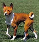
Breeds most commonly reported to be affected by lymphangiectasia are the Basenji, the Norwegian Lundehund, Wheaten terriers, Yorkshire terriers, Shar-peis, and Rottweilers. There is no reported sex predisposition. A physical exam of an affected animal might reveal weight loss, emaciation, thickened intestinal loops, and secondary findings expected from protein loss, such as ascites or pleural effusion. The presenting sign to a veterinarian, thus, might be respiratory distress from pleural effusion, not necessarily a problem that necessarily points towards the gastrointestinal tract. Ultrasound evaluations of dogs with lymphangiectasia often reveal hyperechoic mucosal striations from the reflected ultrasound waves off of the dilated lacteals.
Necropsy, biopsies or endoscopic examinations of animals affected by lymphangiectasia have many distinct findings, though varying in degree and extent. The dilated lacteals that are filled with chyle can be seen grossly as elevations in the edematous mucosa of affected segments of bowel. The mesenteric lymph nodes are commonly enlarged, white, and also dilated, as are white nodular masses often seen in the mesenteric border adjacent to enteric lymphatics. Grossly, the intestinal walls are thickened, white foci or nodules are present on the serosal surface and mesenteric lymphatics are distended. Histological evaluation usually reveals blunted villi with hypertrophic crypts. The intestinal epithelium is variably attenuated, normal, or spaced far apart. Most characteristically, the lacteals within the villi, and commonly in the mucosa or submucosa, are markedly distended. Macrophage and lipid infiltrations can be seen and correlate with the occasional presence of white nodular masses if seen grossly. Edema of the intestinal wall is usually present.
Neither human nor veterinary medicine has successfully offered a curative therapy for lymphangiectasia other than nutritional modulation of diet. Dietary treatment consists of low-fat and normal caloric intake with albumin or colloid fluid supplementation. Corticosteroid therapy may be helpful if concurrent inflammation is present. Response and prognosis is variable, usually in direct correlation to the extent of the disease. Current research in treating human intestinal lymphangiectasia with long-acting somatostatin analogues may work locally by promoting small intestinal mucosa healing. This treatment is administered alongside the aforementioned dietary therapy.
References:
- Peterson PB, Willard MD. 2003. Protein-losing enteropathies. Veterinary Clinics of North America: Small Animal Practice 33: 1061-1082.
- Filik L, et al.: 2004. A case with intestinal lymphangiectasia successfully treated with slow-release octreotide. Digestive and Liver Disease 36: 687-690.
- Kimmel SE, et al.: 2000. Hypomagnesemia and hypocalcemia associated with protein-losing enteropathy in Yorkshire Terriers: five cases (1992-1998). JAVMA 217(5): 703-706.
- Jubb, Kennedy, and Palmer’s Pathology of Domestic Animals, 5th edition, Vol. 2m Edited by Maxie, M Grant. 2007.
- Kull PA, et al.: 2001. Clinical, cliniopathologic, radiographic, and ultrasonic characteristics of intestinal lymphangiectasia in dogs: 17 cases (1996-1998). JAVMA 219(2): 197-202.
- Clinical Veterinary Advisor: Dogs and Cats, Edited by Côte E, 2007.
- Sutherland-Smith J, et al.: 2007. Ultrasonographic intestinal hyperechoic mucosal striations in dogs are associated with lacteal dilation. Ultrasound 48(1): 51-57.
- Willard, MD, et al. Intestinal crypt lesions associated with protein-losing enteropathy in the dog. J Vet Intern Med (2000) 13(3): 298-307.