 Equine piroplasmosis (EP) is a tick-borne disease caused by the protozoan parasites Theileria equi and Babesia caballi. Also known as equine malaria or horse tick fever, EP can cause acute hemolytic anemia in horses, mules, donkeys, and zebras. Though the causative agents are endemic in most of the world, equine piroplasmosis was eradicated from the US in 1988, a process that took 25 years and 12 million dollars to complete. However, a 2009 outbreak in Texas has stimulated increased interest in the disease.
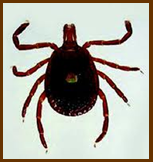
T. Equi and B. caballi belong to the phylum Apicomplexa, order Piroplasmida. The parasites are typically found in tropical and subtropical regions of South and Central America, Africa, Europe, and the Middle East. T. Equi and B. caballi are found in the same geographic locations, and commonly co-infect horses. These agents are transmitted by more than 15 species of ticks within the genera Dermacentor, Hyalomma, and Rhipicephalus. However, key differences in the replication cycles of Babesia and Theileria allow for separate modes of transmission.
B. caballi can persist and multiply in ticks for multiple generations because it is both transtadially and transovarially transmitted. The tick serves as the reservoir host for B. caballi. Placental transmission is not a source of infection with B. caballi. The life cycle of T. Equi is slightly different in that the protozoa do not multiply in the tick and there is no transovarial transmission. Equids are the primary reservoir for T. Equi. In utero infection with T. Equi can result in abortion, stillbirth, or neonatal infection. Some foals can become healthy carriers if they ingest colostral antibodies. This phenomenon is known as premonition and occurs in endemic areas.
Parasitic sporozoites develop and travel to the salivary glands to be transmitted when the tick bites a subsequent host. When an infected tick attaches to a host, the Babesia or Theileria parasite is stimulated to undergo final maturation. Therefore, ticks must remain attached to the host for several days before they become infective. Once introduced into a naïve horse, B. caballi and T. Equi sporozoites enter erythrocytes and develop into merozoites, which infect other erythrocytes. The incubation period for T. Equi piroplasmosis is 12 to 19 days, and 10 to 30 days for B. caballi. Whereas ticks are the biological vector, mechanical transmission can occur iatrogenically via the use of contaminated instruments, needles, syringes, or blood transfusions.
It has been suggested that equids can carry B. caballi for 4 years or more, and the organism could eventually be cleared. However, what actually happens is the parasitemia decreases to undetectable levels. If the equid is immunosuppressed or exercised strenuously the parasitemia level will again increase. During the time the equid is “clear” of infection and not displaying clinical signs, it serves as a reservoir for infection of other animals.
In endemic areas of the world, most horses become infected with piroplasmosis within the first year of life. The clinical signs of infection are variable and nonspecific between the parasites, but T. Equi tends to be more severe due to destruction of nearly 20% of the horse’s erythrocytes. The clinical signs are due to erythrocyte destruction, complement activation, and release of inflammatory mediators. The differential diagnosis for the clinical signs seen with EP include equine infectious anemia (EIA), erlichiosis, liver failure, autoimmune hemolytic anemia, purpura hemorrhagica, and toxicosis (phenothiazine, wild onions, red maple leaves). Exotic diseases in the differential diagnosis include surra, dourine, and African horse sickness.
Inapparent carrier horses have low levels of parasitemia and no overt clinical signs. However, acute disease can occur in carrier animals that become stressed. Clinical infections can be identified as peracute, acute, subacute, or chronic. The rare peracute infection occurs when a naïve horse is introduced to a large number of infected ticks. CNS signs and sudden death may occur due to the massive destruction of red blood cells leading to severe anemia. These sudden death events also occur after strenuous exercise. More often, horses present acutely with anemia, pyrexia, anorexia, recumbency, labored respiration, dehydration, congested mucous membranes, and tachycardia. Sweating with edema of the distal limbs and supraorbital regions is often noted. Icterus, hemoglobinuria or bilirubinuria can be seen in severe cases due to the release of free hemoglobin into the blood. Pneumonia can follow pulmonary edema. Signs of colic, constipation, and diarrhea can occur with intestinal involvement and dehydration. Kidney failure may occur due to inflammation of blood vessels and massive quantities of hemoglobin in the blood stream. Horses with subacute piroplasmosis have a marked general weakness with anorexia, tachycardia, tachypnea, weight loss, intermittent pyrexia, distal limb edema, and a normocytic, normochromic anemia. Urine may be dark yellow; mucous membranes vary from pink to bright yellow. Lastly, chronic cases present with poor performance, weight loss, and mild inappetence. These horses are difficult to diagnose because anemia may be minimal. Mortality rates may approach 50% in some areas of the world.
The severity of gross and histologic lesions at necropsy depends on the degree of anemia, duration of infection, and age of the horse. Marked subcutaneous edema and icterus is often seen. Other lesions include hepatomegaly, splenomegaly, enlarged kidneys, ascites, hydrothorax, and hydropericardium. Petechial hemorrhages are found throughout the body. Histologic lesions include erythrophagocytosis with hemosiderosis of the liver, spleen, and lymph nodes. Congestion and edema of the lungs and centrilobular necrosis of the liver with multifocal hepatitis is often seen. In the kidney, hydropic and fatty degeneration of the tubular epithelium occurs with protein and hemoglobin casts in tubules.
Diagnosis of EP can be based on clinical signs, but because most horses are asymptomatic carriers and because of the similarity to other equine diseases, diagnosis relies on laboratory testing. Examination of a Giemsa-stained blood smear obtained from acutely infected horses can reveal the protozoa in erythrocytes. B. caballi is pyriform and in pairs, whereas T. Equi appears as four pyriform parasites in a Maltese cross arrangement. Horses that recover from EP and become chronic carriers tend to have low-level parasitemia, as do infections with B. caballi. Therefore, it is recommended to use a thick blood smear technique during the acute phase of infection in order to increase the sensitivity.
With inapparent carriers, it is very difficult to find the organism microscopically, so serology is recommended. Compliment fixation testing (CFT) was once the official standard test for equine piroplasmosis; however, it requires the production of large quantities of antibodies and can be affected by natural anticomplementary activity and drug treatments. Therefore, the test frequently yields false-negative results. There is also cross-reactivity between B. caballi and T. Equi. These false-negative results may have allowed the importation of infected horses into the U.S. The indirect immunofluorescent antibody test (IFAT) involves parasite antigens reacting with test sera on a glass slide. IFAT is more sensitive than CFT but is time-consuming and difficult to standardize. IFAT is often used in conjunction with CFT results. The OIE (World Organization for Animal Health) recognizes the IFAT and cELISA tests as the standards for importation. In 2005, the official U.S. import test became cELISA, which detects antibodies to T. Equi and B. caballi. The use of ELISA increased the sensitivity of import testing. Polymerase chain reaction (PCR) is a highly specific and sensitive method of testing for the parasites in blood. Assays have been developed to detect both T. Equi and B. caballi. However, due to high costs, PCR has been limited to research purposes. Care should be taken when dealing with EP blood and diagnostic testing. Some species of Babesia and Theileria can be zoonotic, although B. caballi and T. Equi do not appear to be a major source of zoonoses.
Importantly, EP is classified as a foreign animal disease in the U.S., and it is illegal to treat an equid in this country outside of government programs. Also, because EP treatments have not been proven to be curative, all treatments are considered experimental. Treated animals may be free of signs of infection, but not sterile (and still able to transmit the disease). Treated animals are subject to recrudescence during times of stress or strenuous exercise. Attempts to clear a horse of infection is not recommended in endemic areas because this would cause the horse to be susceptible to reinfection and developing clinical disease. Treatment could potentially lower resistance and cause exacerbation of clinical signs.
There are 5 options for owners of an infected equid in the U.S.: permanent quarantine, export, euthanasia, donation to a research program or enrollment in an ongoing USDA research project to evaluate therapeutic options.
Prevention and control is often quite difficult for tick-borne diseases, but is accomplished by placing EP-positive animals in quarantine, implementing strict isolation protocols on imported horses (especially those from endemic areas), utilizing permethrin-based acaricides to repel ticks, and tick removal. Imported equids should be quarantined for 30-60 days and retested upon arrival into a country. Rodents and other wildlife must be prevented from entering the quarantine area. Other preventative methods are proper disinfection of tattoo and dental equipment, not reusing needles, and not using an EP-positive horse as a blood donor. There is no vaccine currently available for equine piroplasmosis.
EP has become an important topic within the veterinary world. The fall of 2010 brought the World Equestrian Games (WEG) to Lexington, KY. Typically, horses testing positive for B. caballi or T. Equi are not allowed entry into the U.S. However, for the 1984 and 1996 Summer Olympic Games, seropositive horses were granted waivers to enter the U.S. and compete, just not in field events, which would provide the most tick exposure. For the recent WEG in Kentucky, positive horses were allowed to compete in all events. The USDA, KDA, and APHIS evaluated the tick population in Lexington in order to gauge the risk of EP transmission during the WEG. They found the tick population drops significantly in September when the WEG would be taking place. These organizations instituted policies to reduce the risk of transmission such as natural tick barriers, establishment of designated grazing areas (each area would be treated with a tick repellant), individual stables, tick inspection of horses when they re-entered the stables, and acaricidal treatment effective in killing attached ticks. EP-positive horses were shipped from the federally mandated quarantine center of their country to specific predetermined stables in the Kentucky Horse Park, and were then shipped out of the country directly from these stables.
The recent U.S. outbreak of T. Equi began in early October, 2009. This outbreak caused the Indiana Board of Animal Health to implement an emergency rule in January 2011, and again in April 2011 requiring horses to be tested for EP before entering a premise in the state where Thoroughbred or Quarter Horse races are held. This rule includes pony horses and any other equids used on those premises. The emergency rule states that every equid entering the premises must have been tested within the last 12 months and be retested at least once every 12 months. Any equid not meeting those requirements must leave the premise. The blood must be drawn by a licensed and accredited veterinarian. The cELISA must be sent to a National Veterinary Services Laboratory (NVSL) and tested for both T. Equi and B. caballi. Every equid that enters the premise must have an official copy of the laboratory results, including the animal’s identification, date the sample was drawn, and the negative test results. Any equid that tests positive will not be allowed to enter the premise, and must leave if already on the grounds. Purdue’s ADDL is not a USDA-certified lab, but will accept the samples and forward them to a NVSL. The emergency rule is effective through August 2011, after which the permanent rule will be placed into effect. Due to the global nature of the equine industry, equine piroplasmosis will continue to be a prominent disease within the veterinary world.