By Dr. Nelson Bricker, Class of 2011
Edited by Dr. Tsang Long Lin, ADDL Pathologist
Introduction
Viral hemorrhagic septicemia (VHS) is a disease of global concern in fish for aquaculture as well as wild species. Until the last decade, in the United States, the virus had only been detected in coastal regions of the west coast, including Alaska. The discovery of this disease in the Great Lakes region of the United States in 2003 has almost completely frozen the aquaculture industry in this region in an attempt to prevent further spread of the disease by transport of live fish. While that is an important step, the migration of wild fish, movement of water within a watershed, and transmission by fomites may still be important means of viral spread. The high associated mortalities of susceptible fish and wide range of clinical presentations makes this a disease of high concern to people, not only involved in aquaculture, but also sport fishing and environmental welfare. Sensitive and specific means of detection and differentiation of VHS virus are necessary for identifying both clinically affected fish and carriers of the virus in order to accurately assess the spread of the disease as well as effectively control and eradicate VHS.
Virus Characteristics
 VHS virus (Family: Rhabdovirus, Genus: novirhabdovirus) is a single-stranded, enveloped, negative-sense RNA virus. Viral strains are grouped based on genetic similarity and are associated with geographic regions, environments, and fish species. European strains are numbered I, II, and III. The coastal North American train is known as IVa, and the strain found in the Great Lakes region is being classified as IVb. Several differences have been noted in the IVb strain compared to previous forms in terms of pathogenicity for species affected. However, progression and lesions manifest are similar. VHS has historically affected salmonid fish most severely; however, the IVb strain appears to affect other species as well (e.g. walleye, largemouth bass, freshwater drum, gizzard, shad and yellow perch). So far, VHS is still limited to cold-water species, and shows the greatest outbreaks in the winter, when water temperatures are between 3 and 12 degrees C (37 and 54 degrees F). Indoor aquariums, therefore, are likely to never reach temperatures where viral replication leads to clinical disease. Some koi and other species kept outside may be at risk; however, carp species are generally not affected by clinical disease. While this is obviously a benefit for them, it means that surveillance for carries is still warranted when considering pet fish as a vector for the spread of disease.
Transmission and Epidemiology
VHS virus has the potential to transmit by several methods and can survive in a moist, cool environment. Cohabitation, immersion, intraceoelomic and intramuscular injection, brushing virus on gills, contact with secretory products, and feeding on infected fish have all been experimentally shown to transmit disease. Some fish are more resistant to certain modes of transmission as it has been noted that wild salmonids do not acquire overt disease from VHS-IVb by eating infected fish, but pike have been found to break with the disease by this route. Transmission by cohabitation is generally not considered as significant a factor in wild populations compared to farmed fish, as viral load in water bodies rarely reaches an infectious dose. Wild fish tend to be more resistant to VHS infection due to decreased stress and population density. Mechanical transmission has been implicated from piscivorous birds, fishing, and aquaculture equipment. Aquaculture effluent is considered a significant source of viral particles from infected farms that do not treat water properly. Survival in water may be more than a week if temperatures are at or below 14 degrees C. Unfortunately, fish that are infected and survive are lifelong carriers of VHS viruses. Carriers and clinically affected fish shed viral particles mainly by urinary and sex organ secretions. While viral particles are present on eggs for several hours after laying, vertical transmission does not occur. Younger fish appear to be more susceptible to disease and have a greater mortality rate. However, an incubation period of 1-2 weeks means clinical signs may not occur until fish are older than that age. Most fish reported to die from VHS-IVb are actually mature individuals, though experiments using juveniles has shown their susceptibility as well. It is rare to see significant disease in fish greater than six months of age infected with other strains of VHS virus.
Clinical Course
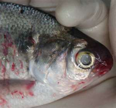
Virus is considered to enter the gills at the level of the secondary lamellae where initial replication takes place. Systemic spread occurs as the virus invades and replicates in the cytoplasm of endothelial cells. The virus is shown to have highest concentrations in the cranial or anterior kidney and spleen compared to other visceral organs and muscle. It has been noted that the carriers of the disease may only have the virus isolated from the brain. Fish may either be acutely affected or develop a low-grade, chronic infection if disease develops at all. Acute disease is characterized by lethargic and anemic fish that develop dark pigmentation of the skin and exophthalmus from retrobulbar hemorrhage and distended coelomes secondary to organomegaly. Hemorrhages are often noted in the skin, eyes, gills and at bases of fins. A leucopenia develops as expected from the high viral loads in hematopoietic organs. Fish may also exhibit behavior change, congregating near water outflow screens in aquaculture facilities. Mortality rate is high in acutely affected fish. Chronic infections show less severe changes and decreased mortality. Hemorrhages are generally less obvious. Hepatic, renal and splenic edema often occur leading to coelomic distension. Skin darkening may still occur, and erythrocyte precursor pleocytosis may occur in the peripheral blood as well as development of a hypoproteinemia. Behavior changes, including a looping swim pattern or darting and spiraling at the bottom of the pond may be seen. Latent infections or carrier animals may exhibit no obvious changes, but can be mildly hyperexcitable and may show a low-grade hypoproteinemia and erythrocyte precursor pleocytosis. Any fish that apparently recovers from an infection of any type is considered a latent carrier for life and may not even shed virus until sexual mature is reached. Epizootics may develop depending on the number of susceptible fish (young fish of a susceptible species) present during high concentrations of virus in the water (spawning season, near infected outflow or contaminated aquaculture facility) under proper conditions (cold temperatures).
Pathology
Lesions associated with VHS in fish generally are vasculitis and hematopoietic tissue necrosis. Distribution of lesions varies depending on the severity of infection. Susceptible fish develop severe acute lesions that progress rapidly. Dark coloration of the skin occurs. Anemia may be noted by paleness of gills and erythropoeitic tissues. Exophthalmia or “pop-eye” may be present in a large number of individuals and is seen secondary to retrobulbar hemorrhage. Hemorrhage is also seen within the eyes, skin, gills, muscles, swim bladder, at the base of fins, in coelomic organs, or into the coelomic cavity. Spleen, liver and kidneys may be congested. Microscopically, liver, spleen and kidney may have cellular vacuolization with pyknosis or karyolysis, intracytoplasmic or intranuclear inclusion bodies. Lymphoid and hematopoietic tissues also undergo necrosis. Chronic VHS is often presented with edema of coelomic organs without obvious hemorrhage. Renal and splenic lymphoid and monocytic precursors appear hyperplastic but can also show focal areas of necrosis. Renal glomeruli may have histologic alterations similar to membranous glomerulopathies of mammals. In addition, any coelomic organ may have focal areas of hemorrhage or necrosis. One study showed that, with the new IVb strain, bass and perch have shown the greatest number of changes in their liver, skin, gills and gonads, while salmonids proved to be most resistant to infection and showed significant necrosis and hemorrhage in their swim bladders. Fish that are carriers of latent infections do not exhibit any lesions associated with the virus infection. Nevertheless, with the large number of susceptible species and expanding range of the disease, any fish species that exhibits similar lesions to those mentioned above should be considered suspect and investigated further.
Diagnostic Testing
Historically, virus isolation and antibody-based testing have been the gold standard for identification of virus particles from tissues. Only minimal problems with cross reactivity between strains were noted. While virus isolation likely remains one of the most important tools in definitive diagnosis of this disease, molecular tools are now available for fast and sensitive detection of virus in populations of fish. In fish screened from the Great Lakes from 2006-2007, the use of quantitative real-time polymerase chain reaction (qRT-PCR) targeting mRNA showed at least three times more sensitive with no false negatives compared to the results from virus isolation by cell culture. Sampling of tissues is important in all cases and tissues with active high viral load will show the best results. Recommended tissues for sampling acute and chronic cases include cranial kidney and spleen with brain being a desirable sample for latent carriers. It was also noted in the studies of the Great Lakes, the results of qRT-PCR correlate well with those of viral load from virus isolation. This technique is useful for monitoring and surveillance of VHS in susceptible populations; however, it is possible that mutations in the virus will create the need for development of new tests in the future. Viral culture for VHS on a large number of cell lines is readily available but requires specific temperature (14-15 degrees C), pH (7.4-7.8), and media for effective growth. Any population testing positive by PCR should also be subjected to virus isolation to confirm strain of virus, track genomic alterations, and confirm the presence of live virus.
Control
The virus reaches highest concentrations when sick fish are present at a high density. Removal and proper disposal of these animals is paramount to reducing infectious load of a waterway. Viral particles may be destroyed with a number of chemical agents, including ether, chloroform, glycerol, formalin and iodophors. Other common cleaners are likely also effective. Viral inactivation has also been noted by use of UV irradiation, heat causing temperatures of 56-60 degrees C, a pH of 3.5 or less, and drying. Vertical transmission does not occur, and decontamination of eggs with iodophors has been described as a method of destroying viral particles without damaging fish embryos. Contaminated effluent from farms should be treated, as these may be a major ongoing source of infectious particles. Decontamination of mechanical vectors such as boats and equipment, as well as movement of live fish between waterways, should be considered as critical control points.
Conclusion
VHS virus is an organism of extreme importance in aquaculture and has the potential to cause severe damage to, as well as significant impact on, wild species as fish populations are placed under high demand from commercial fishing. Currently, the likelihood of the virus invading the pet fish industry is minimal but, as it has been shown extreme variation in species preference for pathogenic effects in new strains, nothing should be completely ruled out for the future. As the virus continues to mutate and invade other waterways, we must be prepared to track its impact. The seasonality and other conditions, which the virus requires for reproduction and pathogenicity, make it difficult to track the periods of latency; however, new molecular techniques should be able to overcome this hurdle in surveillance. As far as the control of VHS in the United States is concerned, control methods instituted by European nations should be considered when developing our own control methods. Several nations have even been able to eradicate the disease. With proper procedures, containment of the organism should be a major short-term goal. To do this properly, early identification of affected waterways, as well as education of those using a water body for commercial or personal reasons, must be a priority. Knowing the characteristic lesions caused by a VHS virus, using sensible clinical awareness and judgement, and applying timely screening for VHS virus by qRT-PCR should allow for rapid identification of possibly infected populations and effective control of VHS in the United States.
- Ammayappan A and Vakharia VN: 2009. Molecular Characterization of the Great Lakes viral hemorrhagic septicemia virus (VHSV) isolate from USA. Virology Journal 6:171.
- Bain MB et al:2010. Distribution of an invasive aquatic pathogen (viral hemorrhagic septicemia virus) in the Great Lakes and its relationship to shipping. PLoS One 5(4):e10156.
- Garver KA et al: 2011. Development and validation of a reverse transcription quantitative PCR for universal detection of viral hemorrhagic septicemia virus. Diseases of Aquatic Organisms 95:97-112.
- Hope KM et al: 2010. Comparison of quantitative RT-PCR with cell culture to detect viral hemorrhagic septicemia virus (VHSV) IVb infections in the Great Lakes. Journal of Aquatic Animal Health 22(1): 50-61.
- http://www.focusonfishhealth.org/ Focus on Fish Health. VHS-Viral Hemorrhagic Septicemia.
- Kim R and Faisal M: 2010. Comparative susceptibility of representative Great Lakes fish species to the North American viral hemorrhagic septicemia virus Sublineage IVb. Diseases of Aquatic Organisms 91(1): 23-24.
- Kim R and Faisal M: 2010. Experimental studies confirm the wide host range of the Great Lakes haemorrhagic septicemia virus genotype IVb. Journal of Fish Diseases 33:83-88.
- Noga EJ: 2010. Fish Disease: Diagnosis and Treatment. 2nd ed. Wiley and Blackwell. Ames, IA.
- Stoskopf MK: 1993. Fish Medicine. WB Saunders Co, Philadelphia, PA.
- VHSV Expert Panel and Working Group: 2010. Viral hemorrhagic septicemia virus (VHSV IVb) risk factors and association measures derived by expert panel. Preventative Veterinary Medicine 94:128-139.