OPP,
or Maedi (which means "shortness of breath in Icelandic), is a slowly
progressive interstitial pneumonia. Visna (which means "wasting" in Icelandic)
is the neurologic form characterized by leukoencephalomyelitis with
demyelination. OPP is the most common clinical manifestation of OvLV infection
in the United States. Other chronic inflammatory conditions associated with
OvLV include polyarthritis and mastitis.
Ingestion of infected colostrum is
the primary route of OvLV transmission. Horizontal transmission by ingestion
or inhalation of aerosolized virus from respiratory secretions or ingestion of
contaminated food or water also occurs, albeit less efficiently, and is
typically associated with close confinement during winter months. In utero
transmission has been reported, but the frequency is unknown. Initial
infection is followed by a latent period of months to years, during which virus
replication is limited and seroconversion is delayed. An important feature of
retrovirus replication is that viral genetic material becomes integrated into
the host cell DNA, resulting in lifelong infections despite production of
specific antibodies. Approximately 20% of sheep infected with OvLV will eventually
develop clinical disease; however, these animals typically do not show clinical
signs until approximately 2 years of age.
Tentative diagnosis of OPP is often
based on clinical signs, including chronic afebrile pneumonia with progressive
respiratory failure and loss of body condition despite a good appetite.
Expiratory dyspnea, abdominal breathing, and severe tachypnea (up to 80-12
breaths/min) may be observed. Pregnant ewes often give birth to small or weak
lambs. Other clinical signs include chronic polyarthritis and mastitis ("hard
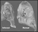
bag"). At necropsy, the lungs have a rubbery consistency, fail to collapse,
and may be 3-4 times normal weight. The basic microscopic lesion in all
affected tissues, including lungs, mammary gland, and central nervous system,
is lymphocytic interstitial inflammation accompanied by formation lymphoid
nodules with germinal centers. OvLV is tropic for mononuclear phagocytes, and
persistent activation of macrophages causes chronic stimulation of the immune
system, resulting in the lymphoid hyperplasia and follicle development observed
in various tissues.
Serologic and molecular-based
diagnostic tests for OvLV are available. Serologic tests such as agar gel
immunodiffusion (AGID) and enzyme linked immunosorbent assay (ELISA)
demonstrate the presence of virus-specific antibodies in serum. AGID is the
most commonly used serologic screening test, but has a lower sensitivity for
detection of antibodies than ELISA-based tests. In experimentally infected
animals, ELISA tests were able to detect seroconversion earlier than AGID. The
PCR test detects proviral DNA in whole blood or tissue samples. The PCR test
is able to detect infected animals before they mount an antibody response, but
is more costly than the serology-based diagnostic tests.
OPP is a chronic, progressive
disease for which no effective treatments or vaccines are available. Control
and prevention programs are paramount. Periodic AGID or ELISA screening tests
are recommended to identify infected individuals in the flock. Lambs may be
removed from infected mothers at birth and raised in separate flocks.
Preferably, these lambs should be fed colostrum and milk from certified
OPP-free ewes. Colostrum from infected ewes should be heated at 56ºC for 60 minutes
and milk should be pasteurized. Alternatively, seropositive ewes may be culled
from the flock. Total herd replacement or annual purchase of OPP-free
replacements should also be considered in lamb-producing flocks. The most
recent National Animal Health Monitoring System (NAHMS) sheep survey in 2001
reported that 24.2% of sheep from the 3,210 operations surveyed nationally were
seropositive for OvLV using the ELISA test. These figures were slightly higher
in the central region, in which 24.4% of sheep tested positive and 46.6% of the
operations surveyed had one or more seropositive animals.
-by Morgan Hennessey, Class of 2006
-edited by Dr. Kim Maratea, ADDL
Graduate Student
References
-
Blacklaws BA, Berruita E,
Torsteinsdottir S, Watt NJ et al: 2004. Transmission of small ruminant
lentiviruses. Vet Microbiol 101: 199-208.
-
Cutlip RC, Lehmkuhl HD, Schmerr
MJ, Brogden KA: 1988. Ovine Progressive Pneuropneumonia (Maedi-Visna) in
Sheep. Vet Microbiol 17: 237-250.
-
DeAndres D, Klein D, Watt NJ,
Berriatua E, et al: 2005. Diagnostic tests for small ruminant lentiviruses.
Vet Microbiol 107: 49-62.
-
Pepin M, Vitu C, Russo P, Mornex
JF, et al: 1998. Maedi-Visna virus infection in sheep: A review. Vet Res 29:
341-367.
-
Peterhans E, Greenland T, Badiola
J, Harkiss G, et al: 2004. Routes of transmission and consequences of small
ruminant lentiviruses infection and eradication schemes. Vet Res 35: 257-274.
-
Petursson G, Matthiasdottir S,
Svansson V, Andresdottir V, et al: 2005. Mucosal vaccination with an
attenuated Maedi virus clone. Vaccine 24: 3223-3228.
-
Petursson G, Hoff-Jorgensen R:
1990. Maedi-Visna and related diseases. Kluwer Academic Publishers, Boston, MA.
-
Preziuso S, REnzoni G, Allen TE,
Taccini E, et al: 2004. Colostral transmission of maedi visua virus: sites of
viral entry in lambs born from experimentally infected ewes. Vet Microbiol
104: 156-164.
-
USDA: APHIS: VS: National Animal
Health Monitoring System: Sheep 2001, April 2003.
|