FINAL DIAGNOSIS: Poxvirus Infection in a Prairie Dog
History: A 12 week-old female prairie dog was euthanized after presentation to the referring veterinarian with respiratory distress. It was then submitted to the Animal Disease Diagnostic Laboratory at Purdue University to determine the specific cause of the clinical signs since deaths of prairie dogs with similar clinical signs had occurred where this animal was bought.
Gross Findings: Gross lesions included numerous variably sized, round, discrete ulcers on the tongue and hard palate, dark red consolidation of both cranial and right middle lobes of the lungs, affecting approximately 40% of the pulmonary parenchyma, and small (<3 mm), white, firm, slightly raised, plaque-like lesions sparsely distributed throughout the wall of the gastrointestinal tract.
Histopathologic Findings: The main microscopic lesions included severe multifocal to coalescing necrotizing bronchopneumonia, with vasculitis and poorly defined eosinophilic inclusions either within the cytoplasm of cells presumed to be of histiocytic or fibroblastic origin or freely scattered throughout the necrotic debris. Multifocal necrotizing lesions, often accompanied by myxoid edema, were also present in sections of nasal turbinates, trachea, thymus and adjacent brown fat, tracheobronchial lymph nodes, lips, tongue, esophagus, stomach, jejunum, cecum, colon, liver, kidney, adrenal gland, vagina, vestibule, female accessory genital glands, conjunctiva, and cornea.
Additional Tests: Ultrastructural examination of lung and intestinal tissue revealed scattered aggregates of immature and mature, non-enveloped, to 320 x 200 nm
viral particles located within the cytoplasm of degenerating cells. The immature viral forms were semicircular to round and had a granular matrix, whereas the mature virions were oval or brick-shaped, with an electron-lucent core and two lateral bodies surrounded by an outer membrane. The morphology of the virions was consistent with poxvirus. Specimens of selected tissues were submitted to the Centers for Disease Control and Prevention (CDC) to confirm the presumptive diagnosis of monkeypox infection. Laboratory evaluation of these tissues is in progress.
Discussion: Monkeypox virus, a member of the orthopoxvirus genus, appears to be enzootic among wild mammals in the west and central African rainforest, where the principal reservoirs are thought to be squirrels and other rodents. Despite the name of the virus, primates are infected only accidentally through direct or close contact with infected reservoir hosts. The disease is usually transmitted to humans from rodents and primates through a bite or contact with the animal's blood. The infection with this virus causes a vesicular and pustular rash similar to, but usually milder, than smallpox. The incubation period is approximately 12 days, and the death rate among infected humans in Africa has ranged from 1-10%. In primates, monkeypox should be considered in any outbreak of a systemic febrile illness that involves a skin rash. Rashes can be severe and generalized in some species. Dyspnea caused by pneumonia may develop in severe cases. Concurrent bacterial septicemia might be present.
The diagnosis is confirmed by histological and electron microscopic examination of tissues, by serological tests (ELISA and hemagglutination inhibition test), and by virus isolation; however, the characteristic lesions on inoculated chicken chorioallantoic membrane and the large eosinophilic intracytoplasmic inclusions typical of poxvirus infections may not be seen with monkeypox virus infection.
Immunohistochemistry, Western blot, and polymerase chain reaction (PCR) tests for monkeypox viral antigen detection are now available.
In the United States, the disease was reported in early June 2003 among several residents of the Midwest who became ill after having contact with sick pet prairie dogs and, in one case, a rabbit. As of 8 July, 2003, a total of 71 cases of monkeypox had been reported to the CDC from Wisconsin, Indiana, Illinois, Missouri, Kansas, and Ohio; these included 35 (49%) cases laboratory-confirmed at CDC and 36 (51%) suspect and probable cases under investigation by state and local health departments. Trace-back investigations have determined that all 35 confirmed human cases of monkeypox were associated with prairie dogs, which appeared to have been infected through contact with Gambian giant rats and dormice that originated in Ghana. Laboratory tests have demonstrated the presence of monkeypox virus in several rodents that died unexpectedly without exhibiting characteristic signs of monkeypox that originated from a shipment from Ghana on 9 April 2003. This outbreak underscores the potential threat to animal and public health by introduction of exotic species.
-by Dr. Ingeborg Langohr, ADDL Graduate Student
 |
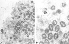 |
Lung, prairie dog, poxvirus infection, transmission electron microscopy. 50,000X (A) and 112,500X (B). Aggregates of immature and mature, non-enveloped virions were located within the cytoplasm of scattered degenerating cells. Mature virions had the typical structure of poxviruses, with an outer membrane enclosing a biconcave core and two lateral bodies. |
| |
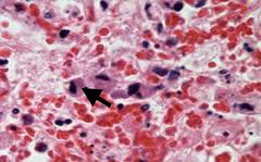 |
Lung, prairie dog, poxvirus infection, 1000X. An eosinophilic inclusion body (arrow) is within the cytoplasm of a degenerating cell presumed to be an alveolar macrophage. |
|
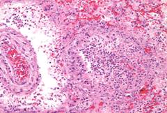 |
Lung, prairie dog, poxvirus infection, 200x. Bronchioles (right), blood vessels (left) and the surrounding pulmonary parenchyma were extensively necrotic. |
|
.