|
Mouser PJ, Ramos-Vara
JA, Vemulapalli R, Scott-Moncrief C. Pathology in Practice. JAVMA 2009 235(10):1153-55.
 Subject: Protozoal encephalitis in a dog Subject: Protozoal encephalitis in a dog
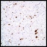
Cerebrum,
dog. Encephalitis. Immunohistochemical detection of Sarcocystis neurona merozoites (individual organisms and clusters) within the inflammatory
infiltrate.
Summary.
An adult dog developed thrombocytopenia, anemia, pyrexia and intermittent
seizures that did not improve with symptomatic treatment. The animal was
submitted to the Purdue ADDL for necropsy. The most significant changes were
present in the cerebrum, which presented asymmetric dilatation of the lateral
ventricles (hydrocephalus). Microscopically, the cerebrum, cerebellum, and
brainstem had multiple foci of inflammation characterized by neutrophils,
macrophages, lymphocytes, plasma cells, and fewer eosinophils. Within the
inflammatory foci there were numerous intra-and extracellular crescent-shaped
merozoites and rare schizonts. The tentative diagnosis was protozoal
encephalitis, most likely produced by Sarcocystis spp. To confirm this
diagnosis, immunohistochemistry for Sarcocystis neurona, Neospora caninum,
and Toxoplasma gondii was performed. Immunohistochemical results were
consistent with a diagnosis of Sarcocystis encephalitis. Molecular analysis
(PCR), using paraffin embedded sections of the affected brain, was performed to
confirm this diagnosis. The amplified genome matched Sarcocystis neurona genome and therefore this diagnosis was confirmed. Sarcocystis neurona is the cause of equine protozoal myeloencephalitis. It has been associated
with cases of encephalitis in dogs. Due to the similarities of microscopic
lesions produced by Toxoplasma, Neospora, and Sarcocystis in the
brain, immunohistochemistry and molecular techniques are necessary to determine
the specific causal agent.
Entire
article available at http://avmajournals.avma.org/doi/full/10.2460/javma.235.10.1153
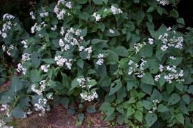
White snakeroot was
growing in the pasture where a heifer died with heart
and skeletal muscle necrosis.
This entire article,
written by Drs. Joshua Webster, Larry Horstman, and Margaret
Miller, can be read in JAVMA 235(7):827-29 or on the
JAVMA website at http://avmajournals.avma.org/doi/full/10.2460/javma.235.7.827
|