|
Contagious Equine Metritis
"A Current Perspective"
Introduction and
History: Contagious Equine Metritis
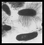
(CEM) is a transmissible venereal disease of equids caused by the gram negative
bacterium, Taylorella equigenitalis. CEM was first diagnosed in Europe
in 1977 but, over the following year, several other countries including the
United States (Kentucky) reported outbreaks. Shortly thereafter, the disease
was eradicated from the U.S. and, until December of 1998, was classified as a
foreign animal disease.
Transmission: Taylorella equigenitalis can be transmitted
directly during breeding of a carrier stallion or mare or indirectly through
artificial insemination or fomites. The highly effective transmission of CEM
can be attributed, at least in part, to unrecognized stallions carrying the
bacteria on their external genitalia. As such, multiple mares may become
infected before the disease is even suspected.
Clinical Signs: There are no reports of stallions infected with CEM
showing clinical signs; however, clinical disease in mares is localized to the
reproductive tract resulting in temporary infertility. Mares can demonstrate
three patterns of infection: acutely infected, chronically affected, or
asymptomatic carriers. Whereas acute infections present with copious, thick,
gray vaginal discharge 10-14 days post breeding, chronic infections have less
vaginal discharge and milder uterine inflammation, but are more difficult to
eliminate. After recovering from acute infections, mares can carry the
bacterium asymptomatically for months. CEM rarely causes permanent infertility
or abortions.
Gross, Histological and
Cytological Findings: During the
initial two to three weeks of infection, the endometrium and cervix are
swollen, edematous, and covered with cloudy gray to white exudates. The
superficial (stratum compactum) and deep (stratum spongiosum) layers of the
endometrium are thickened by edema and infiltrated by neutrophils and fewer
eosinophils which transmigrate into the endometrial epithelium and uterine lumen.
By 14 days post-infection, the edema subsides and the inflammatory infiltrate
consists of lymphocytes, plasma cells, and fewer neutrophils until day 21 when
the inflammation becomes a mixed mononuclear cell population. Inconsistent and
less severe microscopic lesions include suppurative salpingitis and vaginitis.
No lesions are found in the clitoral fossa or clitoral sinus; however,
organisms tend to persit in these locations for longer periods of time and are
therefore common culture sites.
Diagnostics: Reproductive losses associated with CEM could
significantly impact the economic status of the equine industry in the U.S.;
therefore, great efforts have been taken to learn more about diagnosing CEM.
There are currently three tests used to determine if a horse is infected with T.
equigenitalis. These include bacterial culture, serological testing, and
test mating.
Bacterial culture is the
most reliable and accurate way to arrive at a diagnosis; however, challenges
exist with respect to sample handling. A swab of the genitourinary tract may
be taken from the clitoral sinus, clitoral fossa, endometrium, and cervix, or
the preputial folds, urethral fossa, urethra, skin of the penis, and
pre-ejaculatory fluid of the mare and stallion, respectively. Samples are then
placed in liquid Amie's Charcoal medium and refrigerated until it is plated on
chocolate agar. False negatives occur if transit time is prolonged or the
sample is warmed. Despite its reliability, culture is time consuming and
technically demanding, thus newer methods are being developed.
 Serological
testing protocols have been surfacing since the emergence of CEM. The most
commonly implemented test at this time is complement fixation. However, this
test is limited to mares that have produced detectable antibodies to T.
equigenitalis. Polymerase chain reaction (PCR) tests are also used and
have improved efficiency and reliability. In addition, an Enzyme-Linked
Immunosorbent Assay (ELISA) has been designed for detection, but is less
commonly employed. Test mating combines technology from the tests detailed
above. In test mating, a stallion is bred to two CEM-negative mares. The
mares are then tested by culture and serology to check for infection. The
testing procedure requires 35 days before a stallion is declared negative. Serological
testing protocols have been surfacing since the emergence of CEM. The most
commonly implemented test at this time is complement fixation. However, this
test is limited to mares that have produced detectable antibodies to T.
equigenitalis. Polymerase chain reaction (PCR) tests are also used and
have improved efficiency and reliability. In addition, an Enzyme-Linked
Immunosorbent Assay (ELISA) has been designed for detection, but is less
commonly employed. Test mating combines technology from the tests detailed
above. In test mating, a stallion is bred to two CEM-negative mares. The
mares are then tested by culture and serology to check for infection. The
testing procedure requires 35 days before a stallion is declared negative.
Current acceptable
protocols for diagnosis include test mating with culture for stallions and
culture combined with the complement fixation test for mares.
Current status: On December 15, 2008, a quarter horse stallion in
central Kentucky was confirmed to be infected with T. equigenitalis. Fifteen days later, three stallions in Indiana also tested positive for the
bacteria. Epidemio-logical investigations have failed to identify the source
of the outbreak. As of April, 2009, 21 stallions and 5 mares in the United
States have been confirmed positive for T. equigenitalis. From these 26
positive animals, another 960 have been exposed making a total of 986 affected
horses (270 exposed or positive stallions and 708 exposed or positive mares),
covering a range of 48 states (Hawaii and Rhode Island have no links to the
disease). All of the positive and exposed horses were put in quarantine and
underwent testing and treatment protocols. Thus far, 823 (83.5%) of the 986
horses, including all horses from Indiana, completed testing and
treatment protocols and were free of disease.
-by Seth Lundquist, Class
of 2010
-edited by Dr. Chad Frank,
ADDL Graduate Student
References:
-
Acland HM, Kenney RM: 1983.
Lesions of contagious equine metritis. Veterinary Pathology 3:330-341.
-
Croxton-Smith P, Benson JA, Dawson
FL, Powell DG: 1978. A complement fixation test for the antibody to the
contagious equine metritis organism. The Veterinary Record 13: 275-278.
-
Duquesne F, Pronost S, Laugier C,
Petry S: 2007. Identification of Taylorella equigenitalis responsible
for contagious equine metritis in equine genital swabs by direct polymerase
chain reaction. Research in Veterinary Science 1:47-49.
-
Katz JB, Evans LE, Hutto DL, Schroeder-Tucker
LC, Carew AM, Donajue JM, Hirsch DC: 2000. Clinical,
bacteriologic, serologic, and pathologic features of infections with atypical Taylorella
equigenitalis in mares. Journal of the American Veterinary Medical
Association 12: 1945-1948.
-
Katz J, Geer P: 2001. An
enzyme-linked immunosorbent assay for the convenient serodiagnosis of
contagious equine metritis in mares. Journal of Veterinary Diagnostic
Investigation 1:87-88.
-
McKinnon AO and Voss DL: 1993.
Equine Reproduction. London: Lea and Febiger pp 846-848.
-
Timoney PJ: 1996. Contagious
Equine Metritis. Comp Immun Microbiol Infec Dis 3: 199-204.
-
http://www.aphis.usda/gov/newsroom/hot_issues/cem/index.shtml
-
http://www.aphis.usda.gov/publications/animal_health/content/printable_version/fs_ahcem.pdf
|