|

Search
|
|
|
Pulmonary Pneumocystosis
in a Mouse
|
History: An adult female, brown and gray, B6RKO mouse was
submitted dead to the ADDL for necropsy. The history indicated that the mouse
had post-partum bloody vaginal discharge and respiratory distress.
Gross findings: The mouse was in poor body condition. All lung lobes
were diffusely dark red to purple, firm, and oozed bloody fluid on cut section.
Histopathologic findings: Alveolar septa were diffusely thickened by
infiltrating mononuclear cells including macrophages and lymphocytes. Alveoli
were filled with eosinophilic amorphous, granular to flocculent material mixed
with macrophages, neutrophils, sloughed epithelial cells and fewer
lymphocytes. Bronchioles were partially filled with eosinophilic foamy
material, proteinaceous debris and neutrophils. The eosinophilic material
consisted of numerous indistinct, 3-5 microns in diameter, round to ovoid
yeast-like organisms (fungal trophic forms or cysts) with rare pale basophilic
nuclei. Gomori's methanamine silver (GMS) stain demonstrated numerous 3-5
micron in diameter, round to ovoid organisms, consistent with Pneumocystis
murina.
Ancillary testing: No bacteria were isolated from the lung.
|
|
|
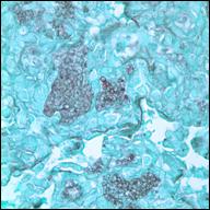
Numerous round to oval cysts are demonstrated within the flocculent intra-alveolar material (GMS, x 100) |
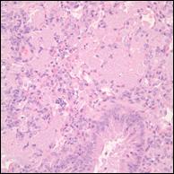
Alveoli are filled with eosinophilic amorphous material containing numerous pale eosinophils to clear, round organisms, which was accompanied by inflammatory leukocytes (H&E, x 40)
|
Discussion: Microscopic lesions with
characteristic intra-alveolar fungal organisms support a diagnosis of pulmonary
pneumocystosis in this mouse. Pneumocystosis in mice is caused by Pneumocystis
murina according to new nomenclature. At one time, all were classified as P.
carinii; however molecular studies have revealed that the genus Pneumocystis contains five species that inhabit different mammalian hosts: P. murina infects mice, P. jiroveci infects human beings, P. carinii and P. wakefieldiae are found in rats, and P. oryctolagi is reported in
rabbits. Other domestic animals are infected by P. carinii. A
lethal pneumonia caused by Pneumocystis spp. is a problem in
immuno-compromised animals, including young dogs, foals, goats, pigs and
laboratory animals as well as humans. Immunocompromised states due to
congenital immunodeficiency, viral infection, chemotherapy, administration of
corticosteroids and other underlying diseases can enhance the growth of Pneumocystis.
The predisposed condition leading to pneumocystosis in this case was not
determined.
Pneumocystis spp.
resides extracellularly in the pulmonary alveoli and, as a fungal organism, a
trophic form (trophozoite) and a cyst (ascus) exist. A trophic form, primarily
the proliferative stage, is 1-4 microns in diameter, uninucleate, irregularly
shaped and thin-walled. A cyst, the reproductive stage, is 5-8 microns,
thick-walled and contains 8 round ascospores. Following inhalation of the
cysts, ascospores are released in the host alveoli and develop into trophic
forms. The infection is initiated by attachment of trophic forms to type 1
pneumocytes with clusters of organisms growing and filling the alveolar lumen.
However, the entire life cycle has not been determined.
Clinical diagnosis of Pneumocystis pneumonia is difficult because
specific alterations in hematological or biochemical parameters or clinical signs
are usually inconclusive. Serology can provide a presumptive diagnosis. Pneumocystis cannot be cultured. Definitive diagnosis is based upon detection of Pneumocystis from respiratory fluid or biopsy samples. Histochemical
stains including GMS, Grocott's, Periodic Acid Schiff and Giemsa, and
immunohistochemistry are useful. Silver stains demonstrate polysaccharide
moieties on cyst walls and intacystic bodies. PAS display the characteristic
honeycombed material of Pneumocystis. Molecular diagnostic techniques
such as in situ rRNA hybridization, DNA hybridization and polymerase
chain reaction (PCR) are developed to identify the specific organisms.
-by Dr. Nozomi Shimonohara,
ADDL Graduate Student
References
-
Caswell JL, Williams KJ:
2007. Pneumocystis carinii. In Maxie MG ed. Jubb, Kennedy and Palmer's
Pathology of Domestic Animals. St. Louis, MO: Elsevier Limited. P. 593.
-
Percy DH, Barthold DW:
2007. Pneumocystis murina Infection: Pneumocystosis. In: Pathology of
Laboratory Rodents and Rabbits. Blackwell Publishing Professional, Ames, IA.
Pp83-84.
-
Keely SP, Fisher JM,
Cushion MT, Stringer JR: 2004. Phylogenetic identification of Pneumocystis
murina sp. nov, a new species in laboratory mice. Microbiology
150:1153-1165.
|
|
|
|
|