Thin strands of fibrin stretched between pleural surfaces and lung lobes. The reticulum was diffusely dark red to purple transmurally, and was well-demarcated from the adjacent normal rumen. The abdomen contained approximately 300 ml of dark red, watery fluid.
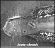
Histologic findings: Glossal mucosa was segmentally ulcerated. Underlying muscle contained broad swaths of coagulative necrosis. Blood vessel walls throughout the tongue were infiltrated by neutrophils and few histiocytes. Severely affected vessels had diffuse loss of endothelial cells, necrotic medial myocytes,and sub-intimal erythrocytes. Adventitia surrounding blood vessels was expanded by clear edema fluid and hemorrhage. Few blood vessels contained hyalinized, eosinophilic material within the vessel wall, indicating fibrinoid change. Fibrin thrombi filled many vessel lumina. Thus, histologic changes were consistent with vasculitis, thrombosis, infarction, ulceration, and hemorrhage.
The reticulum was diffusely ulcerated. Blood vessel walls were infiltrated by neutrophils and few histiocytes; myocytes in the tunica media contained pyknotic and karyorrhectic nuclei. Few blood vessel walls were effaced by fibrinoid material, leukocytes, and necrotic cell debris. Erythrocytes effaced much of the tissue. The changes were consistent with vasculitis, infarction, and hemorrhage. Other histologic changes corresponded with the previously described gross lesions, including fibrinous pleuritis and tissues with multiple petechial hemorrhages.
Ancillary findings: Spleen, lymph node, lung, tonsil, and liver were submitted for Epizootic Hemorrhagic Disease (EHD) virus fluorescent antibody testing and virus isolation. Multiple tissues were submitted in order to maximize the chances of isolating virus. Consequently, EHD virus was isolated from lung, lymph node, and spleen. FA for EHD virus was negative on all tissues.
Discussion: Epizootic hemorrhagic disease of deer is an often fatal disease caused by a double-stranded RNA orbivirus, and transmitted by gnats in the family Culicoides. Epizootic hemorrhagic disease virus is closely related to the orbivirus that causes bluetongue in cattle and sheep. There is serologic cross-reactivity between some serotypes of EHD virus and bluetongue virus. Bluetongue virus can also affect deer, and has been isolated alongside EHD virus from epizootics in North America. Likewise, cattle can become infected with EHD virus and exhibit mild clinical disease which rarely results in death. Lesions in cattle can include oral erosions, lameness/stiffness, and teat erosions. Epizootic hemorrhagic disease virus serotypes 1 and 2 have been implicated in epizootics of deer in the US, with EHD type 2 most often implicated in severe outbreaks. No vaccine is currently available for this disease.
The clinical course of EHD in wild and captive deer is usually rapid, with disease onset approximately seven days after infection. Clinical signs include ptyalism, submandibular swelling, dyspnea, anorexia, or neurological signs. Many deer are simply found dead. Classically, deer found dead near a body of water were thought to have died of EHD as the disease induces body temperatures of up to 106° F. The gross lesions, which typically include petechiae and ecchymoses in multiple tissues, result from systemic vasculitis. Infarcts, with areas of ulceration and necrosis, result from thrombosis of affected vessels. Common locations for infarcts, with subsequent ulceration, include the tongue, oral mucosa, gingival, esophagus, and forestomachs. Commonly, pulmonary edema and fibrinous pleuritis are the only gross lesions observed, both the results of increased vascular permeability.
During the summer of 2007, the Purdue ADDL and Heeke ADDL diagnosed 12 cases of EHD, with 11 positive cases by either FA or virus isolation and 1 suspected case based on gross lesions. Of the 12 total cases, 11 were from White-tailed deer and 1 case was from a beef cow. Cases occurred from August through October. In most cases, numerous deer were affected or found dead, making the actual number of infected deer in Indiana greater than the case load suggests. Submissions came from various locations in both the northern and southern portions of Indiana, and were submitted by deer farmers, veterinarians, hunters, and DNR personnel. Based on ADDL records, the summer of 2007 saw the most confirmed cases of EHD in recent years. When late summer arrives in Indiana, those veterinarians who serve farmed deer should consider EHD as an important differential diagnosis when confronted with seriously ill or dead deer.
-by Dr. Grant Burcham, ADDL Graduate Student
< References:
-
Brown CC, Baker DC, Barker IL: 2007. Alimentary system. IN: Jubb, Kennedy and Palmer’s Pathology of Domestic Animals, ed. Maxie MG, 5th ed., Vol 2, pp 201-203.
-
House C, Shipman LD, Weybright G: 1998. Serological diagnosis of Epizootic Hemorrhagic Disease in cattle in the USA with lesions suggestive of vesicular disease. Annals NY Academy of Sciences. June 29. 849:497-500.
-
Radostits OM, Gay CC, Hinchcliff KW, Constable PD: 2007. Diseases associated with viruses and Chlamydia. In: Veterinary Medicine: A textbook of the diseases of cattle, horses, sheep, pigs, and goats. Elsevier Saunders, Philadelphia, PA.10th ed., p 1305
-
Stallknecht DE et al: 1995. Epizootic Hemorrhagic Disease virus and Bluetongue virus serotype distribution in White-tailed deer in Georgia. Journal of Wildlife Disease 31(3): 331-338.
-
Trainer DO: 1964. Epizootic Hemorrhagic; Disease of Deer. Journal of WildlifeManagement. 28 (2)
|