Adult P. tenuis nematodes
reside in the subdural space of the central nervous system and in the
associated blood vessels and sinuses. The life cycle begins when adult females
lay their eggs in the venous vessels and the eggs hatch in the capillaries
of the lungs. First stage larvae (L1) enter the alveolar sacs and are coughed
up and swallowed. L1 larvae leave the host in the mucus covering of fecal
pellets, then actively penetrate gastropods residing in the pasture. The
larvae molt twice in their intermediate host. Accidental ingestion of the
snails containing infective L3 larvae continues the life cycle. L3 larvae
leave the gastrointestinal tract of the host and enter the central nervous
system in approximately ten days. Larvae develop in gray matter of the dorsal
horn of the spinal cord and migrate to the subdural space 40 days later. In
aberrant hosts, the parasite persists in the parenchyma of the central nervous
system instead of migrating to the subdural space. Disease is caused by
physical trauma to the parenchyma of the central nervous system by developing
and migrating worms.
White-tailed deer are the natural host of P. tenuis; however, other wild and
domestic ungulates have been identified as aberrant hosts and may develop
severe neurologic disease. In response to infection, clinical signs
usually reflect focal, asymmetrical spinal cord lesions and include ataxia,
stiffness, muscular weakness, hypermetria, posterior paresis, paralysis,
head tilt, arching neck, circling, blindness, gradual weight loss, depression,
seizures, and death. Clinical signs generally begin in the hind limbs and
progress to the front limbs. The disease may be acute or chronic, with death
within days to ataxia that lasts months to years.
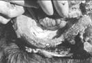
Microscopic lesions include scattered foci of hemorrhagic necrosis. Acute
lesions are characterized by focal parenchymal loss with hemorrhage in and
around the area of injury. Most chronic lesions have no
hemorrhage,
but varying numbers of large, foamy macrophages, some containing gold pigment
consistent with hemosiderin. Around some necrotic foci there can be swollen
axons. The microscopic lesions seen are most compatible with lesions caused
by a migrating parasite.
The use of cerebrospinal fluid for diagnosis of P. tenuis infection is
valuable, especially since hematologic abnormalities are often not
found with meningeal worm infection Eosinophilia in the cerebrospinal fluid is
a common, although inconsistent, finding in aberrant hosts.. Leukocytosis and
vacuolated monocytoid cells are often found. CSF eosinophilic pleocytosis
is not always associated with cerebrospinal parelaphostrongylosis, and other
parasites can cause eosinophilic meningitis in South American camelids.
The only antemortem test for diagnosing P.
tenuis is the Baerman technique, which relies on the
detection of L1 larvae in the feces of infected animals by microscopic
examination. Aberrant hosts rarely shed larvae within their feces, thus this
test is unreliable even when repeated. Experimental ELISA-based
antigen-antibody tests in goats and elk have shown promise but this test is not
currently available. Additionally, an antigen-capture ELISA has
been developed that can detect antigens of P. tenuis in cerebrospinal fluid, but
this test is not commercially available.
The definitive diagnosis of meningeal worm currently requires demonstration of
larval or adult P. tenuis in
the brain or spinal cord of an affected animal at necropsy. Nematodes are
identified on the basis of their size and the following features: lateral cord
cells broader at the base than at the apex, multinucleated intestinal cells,
with no more than two cells per cross section, and polymyarian coelomyarian
musculature. A presumptive diagnosis may be based on clinical signs, exposure,
and response to treatment.
Recommendations for the prevention of meningeal worm infections include the
exclusion of white-tailed deer from llama and alpaca pastures in endemic areas
and clearing thick ground cover to discourage establishment of snail
intermediate hosts. Prophylactic treatment with ivermectin is more effective
against early larval stages because the drug does not cross the blood brain
barrier. Anti-inflammatory drugs are also important for reduction of the
inflammation associated with migrating larvae and the subsequent inflammatory
response to killed larvae. Use of anti-inflammatory drugs is especially
important to prevent the clinical signs from worsening after treatment.
The prognosis of suspected meningeal worm infection is guarded. Some
clinicians suggest that animals that are only able to stand with support have a
much poorer prognosis than those who are able to stand without assistance.
Some animals suffer permanent neurologic damage but remain otherwise
healthy members of the herd.
Meningeal worm infection may be severely debilitating and potentially fatal,
but can be effectively prevented. Simple steps such as routine deworming every
4-6 weeks, minimizing cohabitation with white-tailed deer, 4-6
weeks, minimizing cohabitation with white-tail deer,
and
a clean, dry environment unfavorable for the growth of snails and slugs will
considerably reduce the herd's risk of infection with meningeal worm.
-by
Abby Durkes, Class of 2008
-edited
by Dr. Grant Burcham, ADDL Graduate Student
References:
-
Anderson
DE: Parelaphostrongylus tenuis (Meningeal
Worm): Infection in Llamas and Alpacas-Ohio State University.
http://www.vet.ohio-state.edu/378.htm. Accessed September, 2007.
-
Anderson
RC: 1992. Nematode parasites of vertebrates: their development and
transmission. CAB International, Oxon, United Kingdom. p. 151-208.
-
Baumgartner
W: 1985. Parelaphostrongylosis in llamas. JAVMA 185 (11): 1243-1245.
-
Brown
T, H Jordan, and C Demorest: 1978. Cerebrospinal Parelaphostrongylosis in
llamas. Journal of Wildlife Diseases 14(4): 441-444.
-
Duffy
MS, TA Greaves, NJ Keppie, and MD Burt: 2002. Meningeal worm is a long-lived
parasitic nematode in white-tailed deer. Journal of Wildlife Diseases
38:448-452.
-
Leguia
G: 1991. The epidemiology and economic impact of llama parasites.
Parasitology Today 7: 54-56.
-
Nagy,
DW: 2004. Parelaphostrongylus
tenuis and other parasitic diseases of the ruminant
nervous system. Veterinary Clinics-Food Animal 20: 393-412.
-
Ogunremi
O: 2001. Immunodiagnosis of experimental Parelaphostrongylus tenuis infection in
elk. The Canadian Journal of Veterinary Research 66(1): 1-7.
-
Pugh
DG: 1995. clinical parelaphostrongylosis in llamas. Compendium on Continuing
Education for the Practicing Veterinarian 17: 600-606.
-
Welles
EG et al: 1994. Composition of cerebro-spinal fluid in healthy adult llamas.
American Journal of Veterinary Research 55 (*): 1075-1079.
|