FINAL
DIAGNOSIS: EHD virus infection in a Southern Indiana cow
In August 2007, Heeke ADDL was presented with a 9-year-old Charolais cow with a
7 day clinical history of lethargy, standing in water (suspected fever), poor appetite
and weight loss. A total of 3 cows in this herd of 42 were exhibiting similar
clinical signs, though this was the only cow that had died.
Grossly, the cow had ulcerative and necrotizing rhinitis, stomatitis,
reticulitis, and teat dermatitis, along with pulmonary congestion and edema and
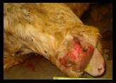
fetal mummification. The epithelium of the nares was markedly reddened and
necrotic and had extensive areas where the epithelium was sloughed. The nasal
turbinate mucosa was diffusely reddened but otherwise grossly unremarkable.
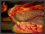
The dental pad and the epithelial ridges of the palate were severely sloughed
and ulcerated. The buccal mucosa had multiple 1-2 mm red shallow erosions,
often surrounding the buccal papillae. The gingival mucosa surrounding the lower
incisors was yellow-gray and necrotic, with hyperemic margins. The tip of the
tongue was dark, dry, and sunken (necrotic), and the epithelium on the ventral
midline of the tongue was white, raised (possible remnant of a vesicle), and
focally sloughed. The epithelium of the lower lip was yellow-gray, dry,
cracked and necrotic. A 3-cm area of the mucosa of the reticulum was gray and
dry (necrotic) with hyperemic margins. Esophagus, rumen, abomasum and
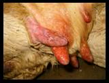
intestine were grossly unremarkable. The skin of the teats had multiple 1-3
mm red erosions and shallow ulcers. The uterus contained a 12x14x30 cm mass of
fetal bones (severely mummified fetus).
Histologically, the cow had erosive and ulcerative stomatitis, nasal
dermatitis, rhinitis, and reticulitis. The oral mucosa had multifocal
full-thickness necrosis and ulceration with numerous superficial bacilli,
marked congestion of the superficial submucosal blood vessels, and mild
perivascular lymphocytic infiltrates. The tongue had a large focal ulcer, with
coagulation necrosis of the underlying vessels. Some blood vessels of the
tongue contained fibrin thrombi, and colonies of thin filamentous bacilli were
present in some of the deeper areas of necrosis. The nasal tip epidermis had necrosis
and separation (sloughing) of the epidermis, marked necrosis of the
superficial dermis, infiltration of degenerating neutrophils into the
superficial dermal papillae, and marked congestion of the superficial dermal
capillaries. The nasal turbinate submucosa was congested and edematous, and
heavily infiltrated with degenerating neutrophils.
Based upon the time of year, the lesions (especially the characteristic dental
pad necrosis and teat lesions), and the presence of an epizootic hemorrhagic
disease (EHD) outbreak in the local whitetail deer population, infection with
EHD virus was strongly suspected. The differential diagnoses included BVD,
IBR, Malignant catarrhal fever, foot-and-mouth-disease and rinderpest. Tissues
were submitted to the Foreign Animal Disease Laboratory at Plum
Island, New York, and no foreign animal diseases were detected. Lip, oral
mucosa, tongue, reticulum, teat, lung, spleen, and lymph node were submitted
for fluorescent antibody (FA) testing and virus isolation. FA tests did not
detect BVD, IBR, EHD virus in any of the tissues, but EHD virus was isolated
from the lung, spleen, lymph node, and oral mucosa. Post-mortem pericardial
fluid was submitted for serologic testing, and was found to be positive for EHD
by agar gel immunodiffusion (AGID). ELISA testing for Bluetongue virus was
negative. A tissue pool was submitted to the National Veterinary Services
Laboratory in Ames, Iowa, and PCR tests were negative for alcephaline
herpesvirus-1 and ovine herpesvirus-2 (the causes of malignant catarrhal
fever). EHD viral RNA was detected in the sample tested by PCR. EHD virus was
then isolated from the pooled tissues by inoculation onto BHK-21 cells and
cattle pulmonary artery endothelium cells, and was determined to be EHD type-2
by virus neutralization testing. No bacterial pathogens were isolated from the
lung and liver. Based upon the characteristic clinical signs and lesions,
and the isolation of EHD virus from the affected tissues, bovine EHD was
diagnosed in this case.
-by
Dr. Duane Murphy, Heeke ADDL
|
|
|
Erosion
of nasal mucosa |
Epithelial
erosions of the
teats |
Erosion
of dental pad and
hard palate |
|