|
Summer 2008 Newsletter
|
|
The four traditional major toxins are alpha,
beta, epsilon, and iota, although C.
perfringens can produce about 15 different toxins
altogether, including enterotoxin, perfringolysin, collagenase, lambda toxin,
hyaluronidase, DNase, neuraminidases, and urease. Enterotoxin is implicated in
cases of human foodborne illness. The more recently identified cpb2, a beta 2
toxin, is believed to be a major contributor to disease in piglets.
The two most common types of C.
perfringens that cause diarrhea in pigs are types A and
C. Traditionally, type C was the most commonly implicated bacteria in
Clostridial diarrhea. Type C is found in extremely low numbers in the normal
GI tract and is typically found in very high numbers in pigs with disease. It
produces alpha and beta toxins. Beta toxin is important in causing a
necrotizing, hemorrhagic disease. Clostridium
perfringens type A is a normal inhabitant of the colon
in pigs and the pathogenesis of disease is not well understood. In a study
from the Netherlands, type A was the most commonly isolated clostridial type
from pigs with diarrhea. Type A produces the alpha toxin, as do all types
of C. perfringens.
However, in experimental studies, the alpha toxin alone did not demonstrate
disease. Both type A and C can produce the cpb2 toxin. In a study by Klaasen,
et al., a large percentage of the diarrheic piglets had C. perfringens isolates
that produced the cpb2 toxin. It is not believed that the cpb2 toxin plays a
role in causing enteritis in piglets with type C and, more importantly, in causing
disease in type A infections. Clostridium perfringens is
a primary pathogen, but it can colonize the intestines after other diseases,
such as transmissible gastroenteritis, coccidiosis, rotaviral enteritis,
and porcine epidemic diarrhea. The organism is transferred by direct
contact between infected piglets and, most importantly, from the sow. The
spores can also persist in the environment and are resistant to heat,
disinfectants, and ultraviolet light. The disease is most common in 3-day-old
piglets, but can affect piglets from 12 hours to 7 days old. Risk factors for
young animals include an immature GI tract, immature intestinal flora, and
the relative lack of trypsin, which can inactivate the beta toxin found in C. perfringens type C.
Clostridium perfringens multiplies to large numbers in a matter of hours, then
attaches to jejunal epithelial cells at villus apices. The toxins produced are
cytotoxic and affect tight junctions and ion transport systems leading to loss
of fluid, electrolytes, and necrosis. Desquamation occurs and the organism
proliferates along the basement membrane. In type C infection, necrosis of the
villous lamina propria with hemorrhage is extensive and the necrotic zone
advances to involve crypts, muscularis mucosa, submucosa, and muscular layers.
In type A infection, attachment and invasion are not as common and induces
a more secretory diarrhea by affecting the tight junctions. The jejunum and
ileum are mainly affected, but both infections can spread to involve
parts of the colon. Clostridium perfringens type
C typically affects 0-7 day old piglets. The fertility rate varies, but is
typically over 50%. The disease can present as peracute, acute, or chronic.
Peracute cases have signs of intense abdominal pain, depression,
weakness, decreased temperature, and bloody diarrhea; death typically occurs
within 24 hours. In acute cases, piglets may survive for 1-2 days after
clinical signs, have reddish-brown diarrhea with gray shreds of tissue debris,
are dehydrated with loss of body condition, and become weak. In
chronic cases, piglets tend to remain active, alert and appetent, but
become progressively thin and dehydrated with intermittent yellow-gray mucoid
diarrhea. In C. perfringens type
A infection, piglets are affected within their first week of life and develop
creamy or pasty diarrhea which may become mucoid and pink. Piglets may
recover, but tend to be stunted.
Features identified during necropsy examination of pigs with peracute and acute C. perfringens type
C include an edematous abdominal wall, intense small intestinal hemorrhage
localized to the jejunum and ileum, emphysema in the wall of the intestine, and
variable peritonitis with bloodstained abdominal fluid. The intestinal
contents are bloody. Mesenteric lymph nodes may be reddened.
Deposition of urate crystals in the kidney is common due to severe hydration.
Chronic cases typically have adhesions between thickened, well-defined affected
areas of small intestine. The mucosa is oftened covered by a tightly adhered
necrotic membrane. Pigs with C.
perfringens type A infection have a flaccid,
thin-walled, gas-filled small intestine with mild mucosal inflammation and
typically no blood.
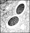
Histopathologically, pigs with C.
perfringens type C enteritis have necrotic jejunal
villi carpeted by large gram-positive bacilli with profuse hemorrhage. The
necrotic area is homogeneous and eosinophilic with scattered pyknotic or
karyorrhectic nuclei and an inflammatory cell infiltrate composed mainly
of neutrophils and some mononuclear cells. Clostridium perfringens type A induces
superficial villous tip necrosis with localized fibrin and gram-positive
bacilli. Clostridium perfringens enteritis
can be diagnosed based on clinical signs, pattern of mortality, and nature of
gross and microscopic lesions. Microscopic lesions with the carpet of large
gram-positive bacilli on necrotic, atrophied villi are pathognomonic. A
definitive diagnosis of differentiation of type can be done by bacteriologic
culture followed by toxin detection or genotyping. PCR methods can also be
used to detect genes for the major toxins. Cultures can be negative in chronic
cases and may yield a mixture of type C and type A organisms. Failure to
demonstrate other agents also supports a diagnosis of C. perfringens infection.
If a type A infection is found, it is very likely that the cpb2 toxin can be
revealed.
Prophylaxis is preferred to treatment in animals with clinical signs. The best
way to control clostridial infections is by vaccination of the sows with a
clostridial toxoid. Currently, most herds are vaccinated with type C toxoid at
breeding or at midgestation and 2-3 weeks before farrowing. A vaccination
program usually eliminates the disease within one farrowing cycle. Ingestion
of adequate colostrums also helps to protect piglets. A type A vaccination is
not commercially available, but can be made via custom biologics. -by
Dr. Lynn Statler, Class of 2008 -edited
by Dr., Pam Mouser, ADDL Graduate Student References: 1Bueschel
DM, et al: 2003. Prevalence of cpb2, endocoding beta2 toxin, in Clostridium
perfringens field isolates: correlation of genotype with phenotype. Vet Micro
94: 121-129 Klaasen
H et al: 1999.Detection of the B2 toxin gene of Clostridium perfringens in
diarrhoeic pigets in The Netherlands and Switzerland. FEMS Immun and Med Micro
24: 325-332. Laohachai
KN et al: 2003. The role of bacterial and non-
bacterial toxins in the induction of changes in membrane
transport: implications for diarrhea. Toxicon 42:686-707. McClane
BA and Chakrabarti G: 2004. New insights into
the cytotoxic mechanisms of Clostridium perfringens
enterotoxin. Anaerobe 10: 107-114. Sawires
YS and Songer JG: 2006. Clostridium perfrin
gens: Insight into virulence evolution and population
structure. Anaerobe 12: 23-43. Songer
JG and Taylor DJ: 2006. Clostridial Infections.
Diseases of Swine 9th ed., Ames, IA. Blackwell Songer
JG and Uzal FA: 2005. Clostridial enteric
infections in pigs. a J Vet Diag Invest 17: 528-536.
|
|
|||||||||||||||||||||||||||||||||||||||