|

Search
|
|
|
FINAL DIAGNOSIS : Fatal systemic canine herpesvirus infection in 2 puppies
|
History:One male and one female 8-day-old puppy were submitted to the Animal Disease Diagnostic Laboratory for necropsy following a history of 7 of 12 puppies in the litter dying within 48 hours.
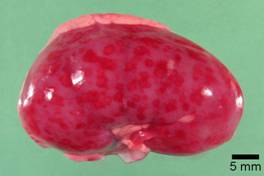
Gross examination:The two puppies had similar necropsy findings. The kidneys contained multifocal hemorrhages apparent from the capsular surface, measuring 2-3 mm in diameter (see Figure 1). The renal parenchyma bulged on cut section, and the hemorrhages extended into the cortex and medulla. The pleural cavity contained transparent, yellow to red-tinged fluid with the quantity ranging from approximately 10-20 mL. The lungs were diffusely mottled red to dark red, slightly firm, and noncollapsing. The livers from both pups had multiple, pinpoint, red foci apparent from the capsular surface and on cut section. |
Pinpoint to coalescing dark red hemorrhages were present on the serosal surface of the small intestine, as well as within the thymus.
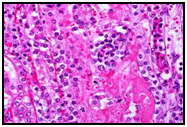
Histopathologic examination: Sections of kidney, lung, and liver from both pups had multifocal parenchymal necrosis. In the kidney, renal tubular epithelial cells were swollen with hypereosinophilic cytoplasm and absent or pyknotic nuclei, and necrotic cell debris filled tubular lumina. Glomerular tufts contained karyorrhectic debris and increased eosinophilic matrix within the mesangium. Endothelial cells lining blood vessels were disrupted, and hemorrhage extended into the necrotic foci.
Basophilic to pale, eosinophilic structures were present within nuclei of tubular epithelial cells and glomerular mesangial cells, reminiscent of intranuclear inclusion bodies. |
|
Figure 1: Gross photograph of one kidney, depicting multiple 2-3 mm, discrete, dark red foci apparent from the capsular surface, corresponding to necrosis and hemorrhage |
In the lung, disrupted alveolar septa were replaced by karyorrhectic debris and fibrin strands. Fibrin, hemorrhage, and foamy alveolar macrophages filled alveoli in affected areas. Pale basophilic to eosinophilic intranuclear inclusion bodies were rarely identified in pneumocytes and bronchiolar epithelial cells at the periphery of necrotic foci. Necrotic hepatocytes in the liver were swollen with hypereosinophilic to vacuolated cytoplasm, and either lacked apparent nuclei or contained pyknotic nuclei. The lesions were not associated with inflammatory infiltrate, but hemorrhage was occasionally present. Rare, viable hepatocytes at the periphery of necrotic foci contained 4-6 micron, lightly basophilic intranuclear inclusion bodies causing margination of chromatin. Nonstaining edema fluid expanded centrilobular connective tissue.
Multifocal necrosis was also identified in the cerebrum, myocardium, adrenal cortex, and small intestinal mucosa.
Ancillary tests:Fluorescent antibody tests for canine herpesvirus were positive on submitted lung samples and on pooled samples of spleen and kidney. In addition, canine herpesvirus was isolated from the lung.
Final diagnosis:Fatal systemic neonatal infection with canine herpesvirus type 1
Comment: The age, clinical history, gross lesions, and microscopic lesions were all characteristic of systemic neonatal herpesvirus infection in this case. The diagnosis was confirmed via fluorescent antibody tests and virus isolation. |
Microscopic identification of intranuclear inclusion bodies can assist in the diagnosis, but inclusions are often rare or absent in canine herpesvirus as compared to herpesviral infections of other domestic species.
-by Dr. Pam Mouser, ADDL Gradaute Student |
|
| Photo From AFIP.org |
|
|
|
|