With regard to S. cruzi, cattle (the intermediate host) ingest the protozoal sporocysts from plant material contaminated with feces of dogs (the definitive host). Initially, two schizogonic generations of the multiplying parasite occur in the vascular endothelium of the infected intermediate host. The merozoites resulting from the second generation schizonts enter the skeletal and cardiac muscle tissue forming sarcocysts over a period of several months. The dog becomes infected by consuming undercooked beef containing the encysted parasite.
Clinical findings: Subclinical Sarcocystis spp. infections are quite common in farm animals; however, clinical disease does not normally occur. When clinical signs are present they are usually non-specific and may include protracted fever, anorexia, decreased milk production, diarrhea, abortion and weakness. Severely affected cardiac muscle may lead to acute death. Acute clinical disease in cattle is referred to as Salmeny disease which is associated with ingestion of massive numbers of infective Sarcocystis sporocysts.
Clinical diagnosis: At present, there is no practical antemortem assay available for the diagnosis of EM. An immunofluorescent assay for S. cruzi antibodies detected significantly higher IgG fluorescence values in bovine carcasses condemned for EM when compared with those which passed inspection, but a distinct cause and effect relationship could not be determined between the parasite and the presence of EM.
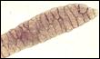
Postmortem diagnosis: EM is most commonly diagnosed during postmortem examination of the affected animal. Gross lesions occur primarily in the heart and striated skeletal muscle and are characterized by focal, firm, greenish gray discoloration. Histologically, the lesions consist of extensive multifocal areas of muscle fiber degeneration and necrosis, with occasional mineralization, atrophy and fibrosis. There is, in addition, a marked inflammatory infiltrate composed predominantly of eosinophils, which accounts for the green appearance of the affected tissues on gross examination.
Pathogenesis: The inability of S. cruzi to consistently produce clinical disease or to cause EM in infected cattle has led many pathologists to doubt that this parasite is the actual cause of the inflammatory condition. The exact pathogenic mechanism of EM is not yet completely understood, but some authors have shown that there may be a relationship between EM and a type-1 hypersensitivity reaction to sarcocysts.
Treatment/Prevention: Since most cases of EM are diagnosed postmortem from subclinical infections, an effective treatment plan is not yet available. Prevention of the condition by preventing the occurrence of sarcocystosis in cattle may be attempted in areas of high incidence of EM; interrupting the life cycle of S. cruzi may, however, prove difficult to accomplish as feces from both domestic and wild canids can easily contaminate feed and water consumed by cattle. Further research is needed to completely establish the pathogenesis of EM, including the possible association with host-dependent, sarcocystic-specific, type 1 hypersensitivity reactions. This may allow for decreased incidence of EM and its adverse economic effects on cattle production.
-by Dr. Jason Hammelman, Class of 2004
-edited by Dr. Ingeborg Langohr, ADDL Graduate Student
References:
-
Aiello SE (ed): 1998. Sarcocystosis. In the Merck Veterinary Manual, 8th ed. Merck and Co., Inc in cooperation with Merial Limited. Pp 880-882.
-
Bowman DD, Lynn RC, Eberhard ML: 2003. Sarcocystis. In Georgis' Parasitology for Veterinarians, 8th ed., Saunders, St. Louis, MO. PP 103-04.
-
Ely RW, Fox JC: 1989. Elevated IgG antibody to Sarcocystic cruzi associated with eosinophilic myositis in cattle. J Vet Diag Invest 1:53-56.
-
Granstrom DE et al: 1989. Type-1 hypersensitivity as a component of eosinophilic myositis (muscular sarcocystosis) in cattle. Am J Vet Res 50: 571-74.
-
Jensen R et al: 1986. Eosinophilic myositis and muscular sarcocystosis in the carcasses of slaughtered cattle and lambs. Am J Vet Res 47: 587-93.
|