Diagnosing Blastomyces dermatitidis In the Small Animal Patient
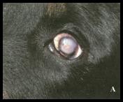
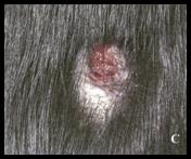
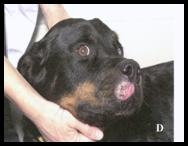
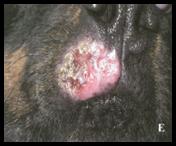
Systemic fungal infections are significant causes of disease because they can gain entry through a single portal and then disseminate to multiple organ systems. A common fungal agent found not only in Indiana, but throughout the Mississippi, Missouri, and Ohio River valleys is Blastomyces dermatitidis. Blastomycosis is most commonly acquired from spore inhalation and colonization of the respiratory tract. Following inhalation, fungal spores are transformed from the mycelial to the yeast phase at normal body temperatures. Dogs and cats can both be affected, but blastomycosis is much more common in dogs. Dogs of any breed or age could become infected if they are in an area with the right environmental conditions for the fungal spores to develop (generally moist, acidic soil rich in decaying vegetation). However, most infected canines have a signalment described as being a young adult, large sporting breed, ranging from 2-5 years of age. There are many clinical signs associated with such a systemic fungal disease. Some of the more common signs that owners notice include anorexia, depression, weight loss, cough, dyspnea, ocular discharge, lameness, and draining cutaneous lesions. Physical exam findings may further include fever, increased lung sounds, lymphadenopathy, uveitis, glaucoma, bone involvement (commonly in the elbow and stifle regions) and cutaneous nodules, papules, or plaques of varying sizes and shapes which can be draining.
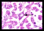
Blastomycosis affects a variety of organ systems; however, the clinical pathology data such as those on complete blood count (CBC) and serum chemistry panel can be very non-specific. One change that can be found, but does not necessarily have to be present, is evidence of chronic inflammation. Most dogs have leukocytosis with mild left shift, monocytosis and lymphopenia. Anemia of chronic inflammation is possible, but many other disease processes can cause this non-specific finding as well. The most common findings that can be found on serum chemistry in an animal with blastomycosis are hypoalbuminemia, hyperglobulinemia, and hypercalcemia. One of the most definitive ways to diagnose blastomycosis is by identifying the yeast organisms retrieved from affected sites by aspirates, impression smears, or biopsy. Cytologic evaluation, which can be performed on body cavity and transtracheal lavage fluid, skin, and organ aspirates, is one of the diagnostic tools that is beneficial when diagnosing blastomycosis. It is a quick and inexpensive way to arrive at a diagnosis. (Modified) Wright's Giemsa stain will adequately stain most fungal organisms in the cytologic preparation. Characteristic features of intralesional B. dermatitidis yeast forms include a thick basophilic cell wall, broad-based budding, are 5-20µm in size, and extracellular location. Cytologic analysis will also provide the type of inflammatory response present, which ranges from pyogranulomatous to granulomatous and can have multinucleated giant cells present. Aspirates of infected lymph nodes, or even exudates or aspirates from the dermal lesions, can contain the fungal organisms. Other common diagnostic procedures performed in dogs with severe respiratory signs include lung aspirate, transtracheal wash, or bronchoalveolar lavage to help find organisms. Bolastomycosis can also be diagnosed through histopathology of tissue samples. There are special immunohistochemical staining techniques that can aid in the identification of blastomycosis in formalin-fixed tissue. Thoracic radiographs, which cah display diffuse or nodular interstitial patterns in the lungs, can help determine how severe blastomycosis infection is, and what course of treatment should be pursued once the organism has been identified.
If you are highly suspicious of blastomycosis, but are unable to find the organism, it may be appropriate to conduct serologic or PCR testing. A variety of serologic tests are available, but agar-gel immunodiffusion (AGID) testing is the most commonly used for B. dermatitidis. This is an antibody test; thus, it can be negative either early in the course of the disease process or in immunosuppressed animals. AGID testing can become negative with treatment or remain positive even with clinical resolution depending on the antibody titer in a particular animal. PCR testing is also available for the diagnosis of blastomycosis, but this testing modality is less useful clinically for this particular fungal agent than for other fungi because of the good quality of the above mentioned diagnostic tests. Culture of B. dermatitidis is dangerous and should only be done by trained personnel.
Treatment of blastomycosis involves long term use of antifungal medications. Other supportive treatments and prognoses are highly variable depending on what organ systems are affected. The best way to maximize the success of treatment is to detect infections early in the disease process.
-by Stacy Amundson-Johnson, Class of 2006
-edited by Dr. Ingeborg Langohr, ADDL Graduate Student
References:
-
Bateman BS: 2002. Disseminated Blastomycosis in a German Shepard Dog. Can Vet J 43(7): 550-552.
-
Dial SM: 2005. Future Diagnosis of Fungal Disease: An Oveview of Molecular and Special Diagnostics. ACVIM Conf Proc.
-
Dial SM: 2005. The Diagnosis of Fungal Disease: Traditional Methods, the Tried and True Diagnostics Revisited. ACVIM Confe Proc.
-
Legendre AM: 1998. Blastomycosis. IN: Infectious Diseases of the Dog and Cat, 2nd ed. Craig Greene, ed. W.B. Saunders Co. Philadelphia, PA. pp. 371-377.
-
Kerl M and Cohn L: 2004. The "F" Files: Case-Based Review of Fungal Diseases. ACVIM Conference Proceedings.
-
Schulman RL and Marks SL: 2005. Systemic Mycoses: Coast to Coast. ACVIM Conference Proceedings.
-
Taboada J and Grooters AM: 2005. Blastomycosis. IN: Textgook of Veterinary Internal Medicine, 6th ed. Stephen J. Ettinger and Edward C. Feldman, eds. Saunders Co. St. Louis, MO pp. 675-680.
|