 |

Search
|
|
|
Final Diagnosis: Lawsonia
intracellularis in a horse
History: A 4-month-old
Tennessee Walking horse filly was submitted dead to the ADDL for necropsy. The
filly was hypothermic and had a history of diarrhea of a few days duration.
The filly became depressed and began exhibiting neurological signs such as head
pressing, mydriasis, and decreased menace response. Fluid therapy, as well as
plasma and flunixin, were administered. Clinical laboratory abnormalities
included hypoproteinemia, azotemia, neutrophilia, lymphocytosis, and several
electrolyte imbalances. The horse was euthanized.
Gross findings:
Segmental areas of jejunal serosa were purple, with prominent serosal and
mesenteric veins. Throughout the jejunum and ileum, intestinal mucosa was
markedly thickened, assuming a cerebriform appearance. |
 |
Multiple areas of
jejunal mucosa were covered with a thin layer of tan fibrin. In severely
affected regions, intestinal wall, including tunica muscularis, was markedly
thickened, measuring over 1 cm in thickness. Circular foci of mucosa, ranging
in size from 1-2 cm, were slightly raised and red; duodenal mucosa was
diffusely red to dark red. The large colon, small colon, and cecum contained
copious amounts of malodorous, dark brown, liquid feces. |
The thorax contained approximately 1-2 liters of clear, straw-colored fluid.
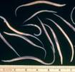
The cranioventral Nematodes consistent
with Parascaris equorum were found within the intestine.portions of both cranial lung lobes were wet and heavy, with
interlobular septa expanded by edema fluid. The abdomen contained
approximately 1 liter of clear, straw-colored fluid.
Histologic findings: Ileal
mucosa was markedly expanded by hyperplastic crypts which contained numerous
mitotic figures and decreased goblet cells. Several crypts were tortuous and
branching. Many crypts were dilated and filled with necrotic debris and
degenerate leukocytes. Large foci of mucosa were necrotic, characterized by
diffuse loss of tissue architecture that extended into underlying submucosa.
Numerous leukocytes, including lymphocytes and neutrophils, expanded lamina
propria replacing some intestinal crypts. Peyer's patches contained decreased
numbers of lymphocytes and karyorrhectic lymphocytes. Submucosa was diffusely
expanded by clear edema fluid. Similar changes were observed within the
duodenum and jejunum, and were consistent with proliferative and necrotic
enteritis.
Alveoli and interlobular septa within the lung were expanded by lightly
eosinophilic material, consistent with pulmonary edema. Clear space surrounded
arterioles within cerebral white matter, giving adjacent neuropil a lacy
appearance.
Histologic changes in the brain were consistent with edema.
Ancillary findings: Two
potential inhabitants of the gastrointestinal system, E. coli and Aeromonas
caviae, were cultured from the intestine. Salmonella culture was
negative. Fecal flotation found numerous ova consistent with Parascaris
equorum.
A section of jejunum tested positive for Lawsonia intracellularis via
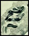
PCR. A Warthin-Starry stain was applied to sections of jejunum and ileum, and
numerous intracytoplasmic bacteria were located in the apical portion of
enterocytes lining hyperplastic crypts.
Discussion:
Characteristic gross and histopathologic lesions, coupled with positive PCR,
were consistent with proliferative enteropathy in this foal. The causative agent
is an obligate intracellular and gram-negative bacteria that is most often
associated with proliferative ileitis in swine. |
|
To date, several species have
reportedly developed disease due to Lawsonia, including horses,
hamsters, dogs, and rabbits. Although the histopathologic diagnosis "prolifer-ative
enteropathy" was made in a foal as early as 1982, the first reported
association between this disease in foals and Lawsonia was made by
authors from Kentucky in 1996. |
Lawsonia- caused
proliferative enteritis occurs sporadically in horses, with both individual
cases and outbreaks on breeding farms. Foals from 3-13 months are most
commonly affected. The most common clinical signs include diarrhea, colic,
weight loss, and ventral or submandibular edema, all of which can be fairly
acute. Common clinical pathologic abnormalities usually reflect a marked
hypoproteinemia due to loss of protein through affected intestine.
Leukocytosis is also a common abnormality. Antemortem diagnosis of this
uncommon equine disease requires exclusion of other, more common, causes of
diarrhea and colic in foals. If proliferative enteritis is suspected after
other causes have been excluded, fecal PCR for Lawsonia intracellularis and serology can be used to aid in diagnosis.
The
above cases differ from previous reports of proliferative enteritis as this
foal rapidly developed severe neurological signs such as head pressing.
Because of previous farm history and recent diagnoses on the same farm, Lawsonia
intracellularis was the likely cause of diarrhea in this foal. Indeed, Lawsonia was confirmed histologically and via PCR. Clinical pathology and gross lesions
were consistent with severe hypoproteinemia; thus, cerebral edema was suspected
as the underlying mechanism for manifestation of neurologic signs.
Histopathologic examination of the brain supported this hypothesis as lesions
suggested cerebral edema. No other cause of neurologic disease was observed.
-by
Dr. Grant Burcham, ADDL Graduate Student
References
-
Brown CC, Baker DC, Barker IK:
2007. Alimentary system. In Jubb, Kennedy, and Palmer's Pathology of Domestic
Animals, ed. Maxie MG, 5th ed. Philadelphia PA, Vol 2, pp 201-203.
-
Lavoie JP et al: 2000. Equine
proliferative enteropathy: a cause of weight loss, colic, diarrhea, and
hypoproteinemia in foals on three breeding farms in Canada. Equine Vet J
32(5): 418-25.
-
McGurrin MK et al: 2007. An
outbreak of Lawsonia intracellularis infection in a standard-bred herd
in Ontario. Can Vet J 48(9): 927-30.
-
Radostits OM, Gay CC, Hinchcliff
KW, Constable PD: 2007. Diseases associated with viruses and Chlamydia.
In: Veterinary Medicine: A textbook of the diseases of cattle, horses, sheep,
pigs, and goats. 10th ed. Philadelphia PA. Elsevier Saunders. p
1305.
-
Williams NM et al: 1996.
Proliferative enteropathy in a foal caused by Lawsonia intracellularis-like
bacterium. J Vet Diag Invest 8: 254-56.
|
|
|
|
|