|

Search
|
|
|
Transmissible Gastroenteritis in
Swine
|
Etiology:
Transmissible gastroenteritis (TGE) in swine is known to be one of the most
significant diarrhea-produceing diseases in young pigs. The causative agent,
TGE virus (TGEV) belongs to the genus Coronavirus of the family Coronaviridae.
TGEV is very stable when stored frozen, but labile at room temperature or
above. TGEV is vulnerable to sunlight and various disinfectants such as sodium
hypochrolite or iodines
To understand an overall picture of TGE, it is necessary to describe a deletion
mutant of TGEV, porcine respiratory coronavirus (PRCV). PRCV appeared in the
1980s and has become prevalent worldwide, including the United States. PRCV
replicates primarily in the respiratory tract and, to a minimal extent, in
small intestinal epithelial cells. Usually, no clinical disease is produced
or, uncommonly, mild respiratory disease is observed. PRCV does not produce
clinical enteric disease. PRCV typically forms an endemic infection in
intensely managed swine herds and infected pigs produce neutralizing antibodies
which also neutralize TGEV. Therefore, when pigs already affected by PRCV get
infected by TGEV, these pigs develop a less severe clinical form of TGE.
Epidemiology:
Epidemiologically, TGE can be classified as epidemic or endemic form.
Epidemic TGE occurs in herds in which most of the pigs are
TGEV/PRCV-seronegative and susceptible, and is observed most often in winter.
As a reservoir during summer months, non-porcine hosts (e.g. cats, dogs, birds)
or mechanical vectors (e.g. houseflies) have been postulated. Morbidity is
high in this form of TGE and pigs under 2-3 weeks of age tend to show severe
diarrhea and rapid dehydration, which often result in death. The mortality
rate is usually less than 10-20%. Diagnosis of endemic TGE in suckling or
recently weaned piglets can be difficult and should be differentiated from
infection with other diarrhoegenic pathogens such as rotavirus, E. coli,
Clostridium spp. and Isospora suis.
Pathogenesis: The major route
of transmission of TGEV is fecal-oral. The incubation period for TGEV is from
18 hours to 3 days. At initial exposure, the pig swallows the virus which then
travels to the small intestine, binds to receptors and is internalized into absorptive
enterocytes. The virus replicates within enterocytes and then lyses them to
enter the intestinal lumen, resulting in villous atrophy. Intestinal crypts
are spared during TGE and become hyperplastic, therefore, secretion continues.
However, absorption is partially impaired by enterocyte lysis and villous
atrophy (malabsorption). At the same time, osmolarity in the intestinal lumen
increases because of the presence of undigested material (maldigestion), which
results from decreased enzymatic activity in the damaged intestinal mucosa.
This increased osmolarity causes a pull of fluid into the intestinal lumen.
Eventually, malabsorption and maldigestion lead to net increase in intestinal
mucosal secretion and clinical manifest diarrhea. The ultimate cause of death
is likely associated with dehydration and metabolic acidosis.
TGEV impacts younger pigs more because their enterocytes are not able to be
replaced as quickly as those of an older animal. Another reason of the higher
morbidity/mortality in younger pigs is that the compensatory fluid absorption
takes place in the large intestine of older pigs, compared with younger pigs.
Clinical signs: In the epidemic
form of TGE, typical clinical signs include transient vomiting, watery, yellow
diarrhea which may contain undigested milk, weight loss, dehydration, and high
morbidity/mortality, especially in pigs less than two weeks of age. One of the
most notable signs is the smell of the diarrhea - foul steatorrhea (excess fat
in feces) due to maldigestion. Many pigs older than three weeks of age will
survive but are likely to remain stunted. Growing and finishing pigs with
epidemic TGE may show inappetance, diarrhea, agalactia, or vomiting of variable
period of time.
Clinical signs for endemic TGE are similar but are less severe than those seen
in seronegative pigs of the same age.
Lesions: Gross examination of
carcasses of pigs that have suffered from TGE will reveal evidence of
dehydration (sunken eyes, increased skin turgor) and diarrhea (soiled perianal/perineal
skin with watery material, lack of formed feces in the large intestine). The
small intestine will have very thin, translucent walls with congested
mesenteric vessels. Mesenteric lymphatics may be devoid of chyle, since there
is malabsorption of fat in the small intestinal mucosa. The stomach and small
intestine may contain a milk curd and bile-stained fluid, respectively. The
small intestine will show villous atrophy if the mucosa is examined by a hand
lens.
The most striking microscopic lesion is severe villous atrophy evidenced by
decrease of villus-crypt ratio (about 1:1 in affected pigs, 7:1 in normal pigs)
in the jejunum and, less commonly, in the ileum. Inflammatory reaction is
usually minimal. Compensatory hyperplasia of crypts develops. Villi may be
lined by attenuated epithelium with irregular nuclear polarity and indistinct
brush border. Since lesions can be patchy, multiple sections should be
examined. Although rotavirus infection can also cause villous atrophy, it is not
usually as severe or extensive as in TGE.
Diagnosis: If TGE is suspected
based on clinical signs and macro-/microscopic lesions, the diagnosis of TGE
can be confirmed by 1) detection of viral antigen, 2) detection of nucleic
acid, 3) microscopic detection of virus at high magnification by electron
microscopy, 4) isolation and identification of virus, and/or 5) detection of a
significant titer increase for TGE. Methodologies apply to each testing are
listed below. Importantly, because of overlapping of clinical signs and
lesions among different etiologies responsible for porcine diarrhea, a combination
of different test methods provides more accurate diagnosis than when a single
test methodology is employed. Please contact the pertinent section of ADDL or
visit our website(www.addl.purdue.edu) for more detailed information regarding methodology, recommended
tissue and timing of sampling, price, etc.
-
Detection of viral antigen: fluorescent antibody assay (FA), immunohistochemistry
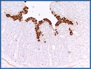
(IHC), Figure 1.
-
Detection of nucleic acid: reverse-transcriptase polymerase chain reaction
(RT-PCR)
-
Microscopic detection of virus: electron microscopy (EM)
-
Isolation and identification of virus: cell culture
-
Detection of a significant antibody response: serology
With regard to serology testing, the diagnosis may be complicated due to
antigenic similarity between TGEV and PRCV. A blocking enzyme-linked
immunosorbent assay (ELISA) can overcome this shortcoming and differentiate
between these two viruses. In order to determine whether endemic TGE or PRCV
is prevalent in a herd, serum samples from pigs of 2-6 months old can be tested
for presence of antibodies. At this age, maternal antibodies should be absent,
thus the positive results suggest endemic TGEV or PRCV. Evaluation of the
results of serology testing always requires careful comparison of disease
history and serological status of the herd.
|
|
|
Figure 1.
Small intestine, pig. Enterocytes at the villous tips show strong cytoplasmic
immunoreactivity to anti-TGEV antibodies. Villous atrophy and crypt
hyperplasia are observed. Immunohistochemistry, hematoxylin counter stain,
200X
|
|
Treatment: The treatment of
choice is supportive care, although it may not be practical under farm
conditions. It is recommended to provide pigs with warm (above 89°F), dry, and draft-free environment. Make water,
nutrient, or rehydration solution accessible because intestinal absorptive
mechanism is intact despite severe damage to the epithelium. Oral fluid intake
will help alleviate severe dehydration especially in pigs that are infected at
older than 3-4 days of age. For 2- to 5-week old pigs, antibiotic therapy may
be helpful if there is concurrent infection by bacterial pathogens.
Prevention and control: In
order to prevent TGE from entering into a sero-negative herd, it is important
to acquire new animals from a TGE-free source which are also sero-negative.
The incoming animals should be quarantined for 2-4 weeks and tested negative
for TGEV before they are introduced into the new herd. Maintenance of a
TGEV-negative herd is based on disciplined biosecurity. Structure of buildings
should exclude all potential animal vectors (rodents, cats, dogs, birds, etc).
Human traffic should be minimized and shower-in shower-out facilities are
ideal. Other fomites (tracks, tools, supplies, etc.) should be carefully
monitored. For more information, please refer to chapter 68 of Diseases of
Swine (reference #5).
The basis of control of TGE in infected herds is to allow the sows to acquire
IgA and continually pass along immunoglobulin-A (IgA) in their milk to provide
passive immunity to the neonatal/suckling pigs. IgA secreting lymphocytes
primarily result from uptake of viral antigens into the M cells in the Peyer's
patches of the intestine rather than from lymphocyte stimulation by a
parenteral vaccine source. Active, protective immunity develops after infection
by virulent TGEV and lasts 6-18 months. Parenteral vaccines typically result
in mainly IgG and IgM production. IgA is stable within the gastrointestinal
tract while IgG and IgM are both destroyed by the digestive process. Since
TGEV is transmitted orally and it targets the enterocytes, it is important to
have IgA functional within the intestinal lumen.
In the event of a TGE outbreak in a naïve herd with no vaccination history, a
later epidemic in the farrowing house appears inevitable. It is important to
begin control at the pre-farrowing level. If sows are two weeks or less to
farrowing, it is important to farrow them off-site or in a non-TGEV exposed
area and keep the young pigs free from exposure for at least three weeks. If
the sows are greater than two weeks away from farrowing, the "feed-back" method
may be used to advertently expose the sows to TGEV and boost their IgA
production which will be passed along in colostrums and milk. To accomplish
this, the small intestines from acutely affected young pigs will be ground up
and fed back to the sows.
In the case of endemic TGE, in addition to all-in/all-out flow and farrowing in
non-TGEV exposed units if available, it is important to use a live attenuated
vaccine to boost immunity of sows prior to farrowing in order to provide a
longer duration of passive immunity to the neonates. This will likely decrease
the morbidity and mortality. The key to the parenteral vaccine boosters and
maintaining immunity is the previous exposure to virulent TGEV. Parenteral
vaccination with an attenuated vaccine in a sero-negative pregnant sow may not
significantly raise IgA levels and thus will not initiate adequate passive immunity
for the neonatal pigs. Finally , some research indicates that multiple
exposures of sows to PRCV resulted in high anti-TGEV IgA in milk and provided a
high degree of protection to TGEV challenge.
-by
Dr. Megan Potter, Class of 2007
-edited
by Dr. Ikki Mitsui, ADDL Graduate Student and Dr. Roman Pogranichniy, Head of
Virology/Serology
References
-
Dewey CE, Carman S, Hazlett M, van Dreumel T, Smart NE:1999. Endemic
transmissible gastroenteritis: Difficulty in diagnosis and attempted
confirmation using a transmission trial. Swine Health Prod 7:73-78.
-
Gelberg HB: 2007. Alimentary system - Transmissible gastroenteritis. IN:
Pathologic Basis of Veterinary Disease. 4th ed., McGavin MD,
Zachary JF eds. Mosby-Elsevier, St. Louis, MO. p. 375.
-
Moeser AJ, Blikslager AT: 2007. Mechanism of porcine diarrheal disease. JAVMA
231:56-67.
-
Murphy FA, Gibbs EPJ, Horzinek MC, Studdert MJ: 1999. Coronaviridae. In:
Veterinary Virology, 3rd ed. Academic Press, San Diego, Ca. pp
495-508.
-
Saif LJ,
Sestak K: 2006. Transmissible gastroenteritis and porcine respiratory
coronavirus. In: Diseases of Swine, 9th ed. Straw BE, Zimmerman JJ,
D'Allaire S, Taylor DJ eds. Blackwell Publishing, Ames, IA. Pp 489-516.
|
|
|
|
|