FINAL DIAGNOSIS: Pituitary abscess syndrome in a sheep
History: A 7-8 year old, spayed Suffolk laboratory ewe was euthanized after presentation to the referring veterinarian with acute recumbency, fever, and dysphagia.
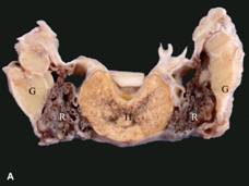
Gross Findings: The skin and auricular cartilage of the left pinna were markedly distorted by a large (~5x1.5 cm) abscess. The ipsilateral middle and inner ear were grossly unremarkable. Viscous green exudate was noted within the pituitary fossa, surrounding the pituitary gland and extending along the caudal fossa of the skull.
Histopathologic description: Extensive coagulative and liquefactive necrosis accompanied by variable neutrophilic infiltrate effaced predominantly the pars distalis of the pituitary gland. Numerous bacterial colonies were present within the affected areas. Additional microscopic alterations included mild chronic otitis externa with focal abscess formation in the left ear, and chronic suppurative perivasculitis affecting the carotid rete-cavernous sinus complex and the cerebral leptomeninges.
Comments: Gross histologic lesions of necrosuppurative hypophysitis (inflammation of the hypophysis or pituitary gland), with intralesional bacterial colonies, were consistent with those seen in pituitary abscess syndrome. This syndrome is an uncommon condition that has been described primarily in humans, cattle, goats and sheep, as well as rarely in horses. The condition is usually fatal. Although reports in the literature usually involve mature (> 2-year-old) animals, the condition has been observed in young cattle after application of nose rings or plastic nose devices to control suckling. Males, both intact and castrated, are apparently more at risk of developing the pituitary abscess syndrome. The aggressive head-butting behavior of bulls, rams, and bucks, leading to sinusitis or cranial trauma with secondary sepsis, might be a contributing factor. The common practice of ringing bull's noses can also be a source of sepsis. Cases are usually sporadic, and even during outbreaks, their occurrence is low (<2% of animals at risk).
Clinical signs may appear suddenly; the course ranges from one day to several weeks. Clinical presentations and histories are highly variable, precluding the description of a classic case. Nonetheless, common clinical findings in affected animals are depression, anorexia, head pressing, exophthalmos, abnormal stance and recumbency; the most common abnormalities noted at neurologic examination are those associated with cranial nerve functions, primarily with ocular function and dysphagia. Cranial nerve deficits are usually asymmetrical and progressive. It is believed that the pressure caused by the enlarging abscess and its extension laterally or dorsally results in damage to adjacent cranial nerves and their nuclei. Bradycardia might be evident, most likely due to compression of the hypothalamus followed by increased vagal tone. A history of infectious disease occurring one to three months before the onset of the neural signs might be reported.
Cerebrospinal fluid analysis is the most rewarding ancillary diagnostic aid. As in most bacterial diseases of the central nervous system, a predominantly neutrophilic pleocytosis with elevated total protein is evident. Definitive diagnosis is made at necropsy since the abscess is usually apparent on gross examination of the brain. In some cases, there is also the presence of osteomyelitis of the basisphenoid bone, single or multiple brain abscesses, and suppurative leptomeningitis at the ventral surface of the brain stem and cervical spinal cord.
The pathogenesis of the pituitary abscess syndrome is uncertain. Direct extension of an adjacent extracranial infection might be a possible cause of the disease. Possibilities include inner ear infection, sinusitis, tooth abscess, infection from a primary cranial fracture, ascending infection through a persistent craniopharyngeal duct and, in the case of horses, guttural pouch empyema. In this particular case, the isolation of identical bacterial organisms (namely Arcanobacterium pyogenes and Escherichia coli) from the ear abscess and the pituitary abscess suggests a causative relationship between these two lesions. The ipsilateral middle and inner ear were grossly unremarkable; the cause of the pituitary abscess by direct extension from the ear abscess cannot be completely ruled out, however, since the middle and inner ear were not further evaluated histologically.
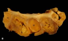
The most likely routes by which bacterial organisms can reach the pituitary gland are arterial, venous, or lymphatic. In ruminants and pigs the gland is surrounded by a complex mesh of intertwined arteries and capillary beds known as the rostral epidural rete mirabile (carotid rete, see figure). This extensive capillary network makes the gland susceptible to bacterial seeding from other chronic sources of infection such as mastitis, arthritis and lung abscesses or pneumonias.
A. pyogenes is the most common isolate from cattle, but a number of other Gram-positive (such as Streptococcus spp., Staphylococcus spp., and Corynebacterium pseudotuberculosis) and Gram-negative (Fusobacterium necrophorum, Bacteroides sp., Pasteurella spp., Pseudomonas spp., Actinobacillus spp.) organisms, as well as Mycoplasma argini, have reportedly been isolated in pure and mixed cultures from pituitary abscesses. These organisms are known to cause chronic suppurative conditions also in other organ systems. This further supports the concept of their circulatory spread.
In addition to dissemination through the blood vascular system, infections in the nasal chamber, paranasal sinuses and ear may spread to intracranial structures by way of the cavernous sinus, a valveless venous system that bathes the pituitary gland and connects with the veins of the extracranial soft tissues of the head. Alternatively, bacteria may gain access to the central nervous system and the pituitary gland through lymphatic channels that communicate with cerebrospinal fluid in the area of the nasal mucosa and the cribiform plate.
Clinically, the differential diagnoses for ruminants with the pituitary abscess syndrome should include listeriosis, bovine herpesvirus 5 infection, polioencephalomalacia, lead poisoning, other brain abscesses, and rabies. Thrombotic meningoencephalitis in cattle, parelaphostrongylosis and coenurosis in sheep and goats, and caprine arthritis encephalitis in goats should also be considered. In horses, differential diagnoses include bacterial meningitis, viral encephalitis (Eastern/Western equine encephalomyelitis, rabies), equine protozoal myelitis, trauma, leukoencephalomalacia, hepato-encephalopathy, and space-occupying masses (abscesses or neoplasia). Clinical presentation and cerebrospinal fluid analysis are usually helpful in differentiating these diseases from pituitary abscesses.
Although infrequently diagnosed, pituitary abscesses can cause signs in ruminants and less commonly in horses that mimic other infectious and toxic neurologic diseases. They should, therefore, be included in the differential diagnosis for central nervous system diseases in these species.
-by Dr. Ingeborg Langohr, ADDL Graduate Student
References:
1. Fernandes CG, Schild AL, Riet-Correa F, Baialardi CEG and Stigger AL: 2000. Pituitary abscess in young calves associated with the use of a controlled suckling device. J Vet Diag Invest 12(1): 70-1.
2. Morin DE: 2004. Brainstem and cranial nerve abnormalities: listeriosis, otitis media/interna, and pituitary abscess syndrome. Vet Clin North Am Food Anim Prac 20(2):243-73.
3. Perdizet JA and Dinsmore P: 1986. Pituitary abscess syndrome. Comp Cont Educ 8(5):S311-8.
4. Reilly L, Habecker P, Beech J, Johnston J, Sweeney C and Hamir A: 1994. Pituitary abscess and basilar empyema in 4 horses. Equine Vet J 26(5): 424-6.
|