During normal hoof growth, attachments between epidermal lamellae and basement membrane are constantly being broken down and then reformed. It is proposed that the cell-cell and cell-basement membrane attachments are released under the influence of active matrix metalloproteinases (MMPs). MMPs have been isolated from normal lamellar tissues and are increased in lamellar tissues of horses affected by laminitis. Recent studies show that bacterial proteases activate MMPs. These results suggest that bacteria can produce potent MMP activators that probably facilitate host invasion.
The enzymatic theory of laminitis based on lamellae MMP activation challenges the alternative view that laminitis develops because of vascular changes to the circulation of the foot. Traditionally, it was suggested that vasoconstriction and compartment syndrome decreased the flow of blood in the lamellar microcirculation to induce ischemic necrosis of epidermal lamellae. A recent study showed that epidermal cell necrosis, intravascular coagulation, and edema were not recognized in sections processed from tissue in the early stages of laminitis. The paper also reported that the vessels in the primary dermal lamella are for the most part fully open without evidence of microvascular thrombi. Further, no abnormalities in the systemic coagulation and fibrinolytic cascades are found in horses with carbohydrate-induced acute laminitis. The gross anatomical appearance of freshly dissected laminitis tissue is one of dryness. Collectively, these results suggest that in the acute phase of laminitis, the enzymatic effect might be the earliest and most influential effect on laminar structures.
The most common causes of laminitis include excessive ingestion of carbohydrates (grain overload), grazing of lush pastures (especially in ponies) and excessive exercise in adult horses. It also may occur secondary to post-parturient metritis, endotoxemia, colic, enteritis, or administration of excess corticosteroids.
Clinical signs of laminitis are often only noted after the disease has progressed and, by that time, the inciting cause may be difficult to ascertain. Once clinical signs of laminitis are present, the clinical course of the disease is typically correlated to the degree of laminar damage. Horses with mild laminar damage have less severe signs and usually respond quickly to therapy. Horses with extensive laminar changes have more severe signs and either respond less quickly or not at all to medical therapy. In general, the acute laminitis affects both front feet, but all four feet could be involved. On palpation, heat may be present over the hoof wall and coronary band. An increased bounding digital pulse is evident. The degree of pain may be reflected by tachycardia, muscle tremors, and sweating. In severe cases, examination is difficult since the horse will often not allow the feet to be picked up and examined.
Obel categorized the severity of lameness by the following criteria: Grade 1, at rest the horse alternately and incessantly lifts the feet, often at intervals of a few seconds; lameness is not noted at a trot. At Grade 2, the horse moves willingly at a walk, but the gait is stilted; a foot can be lifted off the ground without difficulty. At Grade 3, the horse moves very reluctantly and vigorously resists attempts to lift the front foot off the ground. At Grade 4, the horse refuses to move unless forced.
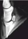
The diagnosis of laminitis is based on clinical signs, physical examination and radiography. Examination with a hoof tester will reveal pain over the sole, particularly at the toe, and tapping on the hoof wall may cause pain. Radiographs should be taken at the first sign of acute laminitis to develop a baseline for continuous radiographic comparison. Early radiographic signs in laminitis include mild bony reaction along the dorsal aspect of the distal phalanx in addition to widening the distance between the distal phalanx and the dorsal hoof wall. This distance should be less than 18 mm in normal horses or less than 30% of the palmer length of the distal phalanx measured from the tip of the bone to its articulation with the navicular bone. Palmar or plantar rotation of the distal phalanx away from the dorsal hoof wall confirms the diagnosis of laminitis. The mean palmar and plantar rotation of the distal phalanx in normal horses is thought to be 0.5 + 1.3 degrees and less than 4 degrees. Digital venogram and vascular perfusion casts have been used to identify perfusion deficits which, if present, usually indicate a poor prognosis.
The goals of treatment are 1) to prevent the further development of laminitis, 2) to reduce the pain or hypertension cycle, 3) to reduce or prevent permanent laminar damager, 4) to improve dermal laminar capillary demodynamics, and 5) to prevent movement of the distal phalanx. Acute laminitis should be considered a medical emergency, and treatment should be initiated as soon as possible, preferably before clinical signs develop. Since circulating endotoxin and infectious processes are found in cases of laminitis, treatment for endotoxemia and sepsis should be attempted. When a horse is suspected of grain overload, one gallon of mineral oil by stomach tube acts as a laxative and tends to prevent absorption of toxic material from the gastrointestinal tract. Recommended treatments include intravenous fluids, parenteral antimicrobials, flunixin meglumine, and hyperimmune serum or plasma. Additional laminitis-preventative measures include the administration of anti-inflammatory drugs, vasodilator, heparin, oral aspirin, and placement of the horse in the stall. Some cases need trimming of the hoof. As for packing of the hoof, a recent study suggests that hot packs used early in the course of the disease may be more beneficial. Non-steroid anti-inflammatory drugs are the cornerstone of most therapeutic regimens.
Predicting the prognosis and survival of horses with acute laminitis can be difficult. One study shows the majority of horses with less than 5.5 degree rotation returned to former athletic function, but those with more than 11.5 degree rotation were no longer able to perform properly, although another paper reported the degree of rotation or distal displacement of the distal phalanx observed on radiographies did not correlate with the outcome of the horses. Based on multiple studies, lameness severity in horses with laminitis likely correlates with the severity or quantity of permanent laminar damage that has, or is likely to, occur.
-by Yasuyuki Usami, ECFVG student
-edited by Dr. Theresa Boulineau, ADDL Graduate Student
References:
-
Aiello SE et al: 1998. Laminitis. In The Merck Veterinary Manual, 8th ed. Philadelphia, National Publishing Inc., 816-818.
-
Brown CM: 2002. Laminitis. In Troy D, ed. The 5-Minute Veterinary Consult Equine. Baltimore: Williams and Wilkins, 602-605.
-
Hunt RJ: 1993. Diagnosing and treating chronic laminitis in horses. Vet Med : 61-64.
-
Hunt RJ: 1996. Diagnosing and treating chronic laminitis in horses. Vet Med 91: 1025-1032.
-
Hunt RJ: 1993. A retrospective evaluation of laminitis in horses. Equine Vet J 25: 61-64.
-
Lindford RL et al: 1993. Qualitative and morphometric radiographic findings in the distal phalanx and digital soft tissues of sound Thoroughbred racehorses. Am J Vet Res 54: 38-51.
-
Mungall BA et al: 2002. Thermolysin activates equine lamellar hoof matrix metalloproteinases. J Comp Path 126: 9-16.
-
Obel N: 1948. Studies on the Histopathology of Acute laminitis. Uppsala, Sweden: Almquist and Wiksells Boktryckteri AK. 1948.
-
Pollitt CC: 1999. Equine laminitis: a revised pathophysiology. Proc 45th annual Conv Am Assoc Equine Pract, 188-192.
-
Stashak TS : 2002. Laminitis. In Stashak TD, ed. Adam's lameness in horses, 5th ed. Baltimore, Williams and Wilkins, 645-664
-
Stick JS et al: 1982. Pedal bone rotation as a prognostic sign in laminitis in horses. J Am Vet Med Assoc. 180: 251-253.
-
Walker M et al: 1995. Radiographic appearance of the feet of mammoth donkeys and the findings of subclinical laminitis. Vet Radiol Ultrasound 36: 32-37.
-
Weiss DJ et al: 1994. Microvascular thrombosis associated with onset of acute laminitis in ponies. Am J Vet Res 55: 606-612.
|