 |

Search
|
|
|
Proliferative Enteropathy in Rabbits |
Proliferative enteropathy is a disease of domestic and laboratory animals; affected species include the pig, horse, dog, rat ,ferret, guinea pig and hamster. The causative agent of proliferative enteropathy is Lawsonia intracellularis,
an obligate intracellular bacterium. The swine industry suffers the most significant impact from this disease. The prevalence of the bacterium and the incidence of associated clinical disease in lagamorphs are currently under investigation.
Lawsonia infection can be common in some rabbit colonies. Lesions are often mild and found as incidental findings in some rabbits at necropsy. Disease rarely results in death; however, severe fatal cases have been noted. Additional viral, bacterial and/or protozoal infections may be responsible for more severe clinical disease. Co-infection with enteropathogenic Escherichia coli has been documented in rabbits with clinical disease.
Epizootology and transmission
Proliferative enteropathy has a worldwide distribution and affects a wide variety of species. Since Lawsonia intracellularis isolates from different species have little genetic variation, intra- and/or interspecies transmission appears likely. Environmental contamination with feces of infected animals appears to be important for transmission of disease, but it is currently unknown how long Lawsonia intracellularis can remain infectious outside of the animal. Animals become infected by consuming fecal-contaminated material. Epizootics of the disease are usually confined to younger animals. Infection occurs often during the post-weaning period, when passive maternal immunity declines. After oral infection, the organisms infect intestinal proliferating crypt epithelial cells and multiply within the apical cytoplasm. There is no evidence of infection of tissue other than intestine. Most animals develop subclinical infection, but shed the bacterium within feces and contribute to environmental contamination. Stressors, such as overcrowding, transport, change in diet, and experimental manipulations have been identified as predisposing factors for clinical infection.
|
Etiology
 |
Proliferative enteropathy is caused by infection with Lawsonia intracellularis, which is an obligate, intracellular, curved rod-shaped, argyrophilic bacterium located within the the apical cytoplasm of infected crypt epithelial cells. |
The means of cellular invasion is still under investigation at this time, but in vitro studies indicate that it involves receptor-ligand mechanisms. Bacteria associate with the cell membrane and are taken up by the enterocyte via an entry vacuole. After lysis of the entry vacuole, bacteria multiply freely within the cytoplasm. Bacterial infection of enterocytes is associated with failure of enterocytes to maturate and increased crypt proliferation. The underlying mechanism is unknown. Animals with subclinical infection do not show clinical signs, but shed the organism within the feces for a limited period of time. Acutely infected animals are lethargic and anorectic, have matted hair coat and watery diarrhea. A common sequela to diarrhea is marked dehydration which may result in cardiovascular collapse. However, enteritis may also cause intestinal intussusception and sudden death.
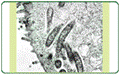
Pathology: On gross inspection at necropsy, affected animals may be emaciated and/or dehydrated. They have proliferative jejunitis and/or ileitis characterized by thickening and corrugation of the small intestinal mucosa and the presence of semi-fluid mucinous contents within lumens of colon and cecum. Microscopically, intestinal crypts are elongated and often branched and lined by multilayered immature enterocytes. The number of goblet cells is decreased. Lumens of crypts may contain cellular debris. Villi are often shortened and blunted and the mucosa commonly has a mixed-cellular infiltration with histiocytes admixed with fewer macrophages, lymphocytes, plasma cells and heterophils and, occasionally, a few multinucleated giant cells. By use of special stains (Warthin Starry silver stain, PAS stain). Agyrophilic and PAS-positive curved bacteria consistent with Lawsonia intracellularis can be demonstrated in apical cytoplasm of enterocytes and within crypt lumens. Intralesional histiocytes may contain intracytoplasmic PAS-positive material consistent with bacterial fragments.
Diagnosis: Diagnosis should be made based on the combination of clinical signs and results of necropsy, histopathology, bacteriology and/or demonstration of intralesional organisms by histochemistry (Warthin Starry silver stain or PAS-stain, see above), electron microscopy, immunohistochemistry and/or polymerase chain reaction (PCR). Bacterial culture and isolation must be performed by use of cultured enterocytes since Lawsonia intracellularis does not proliferate in cell-free media. Electron microscopy can be used to detect the curved bacterial rods consistent with Lawsonia intracellularis within apical cytoplasm of enterocytes. Immunohistochemistry can be performed on formalin-fixed, paraffin embedded tissue of affected intestine to detect intralesional Lawsonia intra-cellularis organisms. In addition, a PCR is available to confirm presence of the Lawsonia intracellularis genome within tissue samples of altered intestine.
Differential Diagnoses: Differential diagnoses for diarrhea in rabbits include Rota and/or Coronavirus, Salmonellosis, antibiotic-associated enteritis, coliform enteritis and intestinal coccidiosis. Coinfections of Lawsonia intracellularis and other bacterial, viral or protozoal pathogens are common and may play an important role in the pathogenesis of the disease.
Treatment: Treatment of ill rabbits consists of symptomatic and supportive therapy. Antibiotics are commonly used to eliminate infection with Lawsonia intracellularis. Isolation of sick animals is advised. Supportive therapy includes administration of parenteral fluids and supplemental heat provided to those animals that become hypothermic.
-by Lou Turchyn, ECFVG Student
-edited by Dr. Sandra Schoeniger, ADDL Graduate Student
References
-
Duhamel, GE et al. 1998: Subclinical proliferative enteropathy in sentinel rabbits associated with Lawsonia intracellularis. Vet Pathol 35: 300-303.
-
Hotchkiss CE et al. 1996: Proliferative enteropathy of rabbits: The intracellular Campylobacter-like organism is closely related to Lawsonia intracellularis. Lab Animal Sci 46: 623-627.
-
Laboratory Animal Medicine. 2002. Fox JG ed., Academic Press, Amsterdam, New York.
-
Hillyer, EV and Quesenberry, KE (eds) 1997: Ferrets, Rabbits and Rodents: Clinical medicine and surgery. W.B. Saunders
-
Percy D and Barthold S (eds) 2002: Pathology of Laboratory Rodents and Rabbits 2nd ed. Iowa State Press.
-
Schauer, DB et al: 1998. Proliferative enterocolitis associated with dual infection with enteropathogenic Escherichia coli and Lawsonia intracellularis. J Clin Microbiol 36: 1700-1703.
-
Suckow MA and Douglas FA (eds): 1997. The Laboratory Rabbit. CRC Press.
|
|
|
|
|