Immunohistochemistry: A Primer for the Practitioner
1. What is immunohistochemistry?
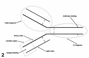
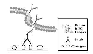
Immunohistochemistry (IHC) uses immunologic and histologic techniques to detect antigens in tissues. The antigen is recognized by a specific antibody (Fig. 1) that is added to the section. The immunologic reaction is visualized under the microscope by adding an enzyme, a substrate to the enzyme and a chromogen, producing a colored reaction (Fig. 2). IHC is a very sensitive and specific technique. For diagnosticians, it is important to colocalize antigens and lesions.
2. Uses of IHC in veterinary diagnostics
Neoplastic and infectious diseases are the main focus of IHC in veterinary medicine. The ADDL IHC Service offers a variety of tests for both infectious and neoplastic diseases. Please contact the ADDL for current tests available and fees.
2.1 Diagnosis of neoplasia. Often, the tissue origin of a tumor cannot be determined with routine histology. Using specific antibodies for different tissues or cells (e.g., cytokeratin for epithelium, vimentin for mesenchymal cells, lymphoid markers, etc); the origin of many tumors can be determined with IHC.
2.2 Detection of micrometastases. Early metastasis can be difficult to detect using conventional histology. IHC highlights the presence of single or small groups of neoplastic cells in metastatic sites. Early detection of micrometastases increases the chances of survival with surgical removal of affected nodes or by modification of the treatment protocol.
2.3 Prognostic markers. Some proteins are expressed in neoplastic, but not in normal mature cells (e.g. embryonal proteins), expressed in neoplastic cells in larger amounts than in normal cells (e.g. cycle-related proteins), or structurally modified in neoplastic cells (mutant p53 protein). These changes may have prognostic significance in specific tumor types. It has been reported recently that the immuno-histochemical detection of KIT protein in mast cell tumors of dogs has prognostic significance. We are testing some of these markers to determine their significance in veterinary cancers.
2.4 Diagnosis of infectious diseases. Detection of antigens of an infectious agent with IHC has etiologic significance. The advantage of IHC over microbiologic techniques is that antigen detection can be correlated with histopathologic changes and thus can confirm the significance of a particular bacterial or viral isolate obtained by other methods. The ADDL offers immunohistochemical tests for infectious diseases of small (feline herpesvirus, Leptospira, canine parvovirus, canine adenovirus, feline leukemia virus, canine distemper virus, etc) and food animals (IBR, BVD virus, TGE virus, Listeria, Cryptosporidium, Neospora, etc).
3. How to submit samples for immunohistochemical testing.
We test samples that have been fixed in formalin, so you do not have to do anything special. Just submit the sample as you would for routine histopathology. Please, do not hold fixed samples in your office longer than 2 days as prolonged fixation may destroy antigens. As soon as you place your sample in formalin, send it to ADDL.
4. Interpretation of results.
Immunohistochemistry facilitates diagnosis of infections and determining the histogenesis and prognosis of neoplasms. A colored reaction (provided that it is specific according to the controls used), indicates the presence of components of the antigen (infectious agent, neoplasm, prognostic marker) sought. Whether this result is significant must be interpreted in the context of the case, as is true for other diagnostic techniques. A careful assessment of the clinical history, lesions and all test results should be made before formulating a definitive diagnosis. Conversely, a negative result by immunohistochemistry does not eliminate the possibility of a particular infectious agent or its potential significance to the case. Due to mutations or other mechanisms, neoplastic cells may modify (upregulate/ downregulate) the expression of proteins resulting in unexpected results. It is important to re-emphasize that immunohistochemistry results, like those obtained by other diagnostic methods, must be supported by clinicopathologic data. Immunohistochemical results should be interpreted by the diagnostician provided that he/she has all the information pertaining to the case.
 In summary, immunohistochemistry is a valuable technique for the diagnosis of infectious and neoplastic diseases of animals. It is sensitive, specific, economical and relatively easy to perform. Although not always considered the"gold standard", it can be as specific as bacterial and virus isolation, provided adequate controls are used. In summary, immunohistochemistry is a valuable technique for the diagnosis of infectious and neoplastic diseases of animals. It is sensitive, specific, economical and relatively easy to perform. Although not always considered the"gold standard", it can be as specific as bacterial and virus isolation, provided adequate controls are used.
-by Dr. Jose Ramos-Vara, ADDL Pathologist, Head of Histology |