|
History: A 2 year old bull elk (Cervus elavus)
was submitted dead for postmortem exami-nation at the Purdue
Animal Disease Diag-nostic Lab. The animal was markedly
emaciated and died following an acute onset of paraplegia.
Gross lesions: Findings at necropsy included marked
infestation by Trichuris sp. Gross examination of
multiple sections of formalin-fixed spinal cord revealed
randomly distributed pinpoint, brownish-red foci both in
the gray matter and white matter between thoracic vertebra
4 and lumbar vertebra 7. These lesions were more severe
and widespread in the white matter. The liver was diffusely
mottled red, and the cut surface revealed the presence of
multiple black fibrotic tracts measuring 0.5-1.5 cm in diameter.
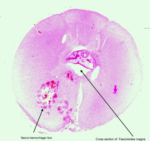
Histopathologic findings: Microscopic examination
of affected segments of spinal cord exhibited irregularly
shaped and-sized multiple foci of necrotic neuropil tissue.
These lesions consisted of acute liquefactive necrosis and
hemorrhage accompanied by moderate numbers of gitter cells,
with occasional randomly scattered eosinophils and neutrophils.
Many swollen eosinophilic axons (spheroids) and swollen
myelin sheaths were present at the margins of necro-hemorrhagic
foci. Blood vessels in the neighboring neuropil had hypertrophic
endothelium and mild perivascular cuffing with lymphocytes
and plasma cells. The leptomeninges in the ventral median
fissure contained large numbers of lymphocytes and neutrophils
and lesser numbers of macrophages, plasma cells, and eosinophils.
In two sections, the central canal was poorly defined, lacked
ependyma, and contained transverse sections of immature
fluke (marita) consistent with Fascioloides magna.
Multiple fibrotic tracts with black pigment were present
in the liver. One of the fibrotic tracts in the liver contained
numerous operculated trematode eggs ranging in diameter
from 40 –60 mm with thick amber to brown shells.
Discussion: Fascioloides magna, the large
American liver fluke or deer liver fluke, is a common parasite
of elk. The flukes are usually confined to the fibrotic
cysts in the liver and, except for causing some liver damage,
are of little clinical consequence. In this case, aberrant
migration of the immature flukes was observed in the spinal
cord and was considered a likely cause of the reported hind
limb paralysis prior to death. The emaciation of this animal
was considered to be due to marked gastrointestinal parasitism.
-by Dr. Alok Sharma, ADDL Graduate
Student
|