 In the summer of 2008, Heeke ADDL was presented with a
one-year old female Boer goat with a two day history of anorexia, "drunken"
staggering, weakness, glass-eyed appearance, and lateral recumbency. Five out
of 20 goats in this herd had died with similar clinical signs. The goats had
been purchased four months prior to the onset of illness, had been treated with
anthelminthic 2 weeks prior to submission, and anthelminthic treatment was
repeated on the day prior to submission. The diet consisted of grass hay and a
grain supplement (Goat Chow). No silage was fed.
In the summer of 2008, Heeke ADDL was presented with a
one-year old female Boer goat with a two day history of anorexia, "drunken"
staggering, weakness, glass-eyed appearance, and lateral recumbency. Five out
of 20 goats in this herd had died with similar clinical signs. The goats had
been purchased four months prior to the onset of illness, had been treated with
anthelminthic 2 weeks prior to submission, and anthelminthic treatment was
repeated on the day prior to submission. The diet consisted of grass hay and a
grain supplement (Goat Chow). No silage was fed.
At submission, the goat was alive but moribund in lateral recumbency. The
animal was in good nutritional condition with adequate muscling and body fat.
Fecal flotation revealed high numbers of Hemonchus contortus ova and
high numbers of coccidial oocysts. The blood had a packed cell volume of 26%.
Following physical examination, the goat was humanely euthanized and
necropsied.
Grossly, the goat had mild meningitis, mild bronchopneumonia, and mild abomasal
hemonchosis. The meningitis was characterized by slight reddening, unusual wetness,
and slight cloudiness of the meninges overlying the cerebellum and the caudal
brain stem. The bronchopneumonia affected only the tip of the right cranial
lung lobe which was red-gray and firmly consolidated. The abomasum contained a
small amount of red-brown watery fluid, and low to moderate numbers of Hemonchus-like nematodes. Grossly, the goat did not appear anemic. The
rumen was filled with finely ground grassy forage and a few fragments of
broadleaves. Intestines were unremarkable and the rectum contained normal firm
fecal pellets. Cytological impression smears of brain stem meninges had
increased numbers of neutrophils, consistent with mild suppurative meningitis.
Histologically, the medulla oblongata, cerebellar peduncle, and midbrain had
multifocal suppurative encephalitis. Scattered mild neutrophilic infiltrates
were also present in the meninges. No histologic lesions were present in the
thalamus and cerebral cortex.
Bacterial cultures isolated Listeria monocytogenes from the brain stem
and Mannheimia haemolytica from the lung. Serologic tests were negative
for caprine arthritis encephalitis (CAE).
Although the clinical history in this case initially suggested parasitism, the
final diagnosis was primary encephalitis listeriosis, complicated by concurrent
abomasal hemonchosis and pneumonic pasteurellosis. Because of similar clinical
presentation, it was assumed by the owner that all six of his dead goats had
died of listeriosis, but the cause of death in the remaining animals was never
confirmed and we cannot exclude the possibility that parasitism contributed to
some of those deaths. The persistence of Hemonchus in the face of repeated
worm medication is indicative of anthelminthic resistance.
Listeria is a common cause of encephalitis in all ruminants, and most large
animal practitioners are familiar with it in cattle. This case illustrates a
number of observations about caprine listeriosis that differ slightly from
bovine listeriosis, and these differences are further examined in an
epidemiologic review of ADDL records below.
ADDL pathology records for the past 5 ½ years (2004-2009) reveal that caprine
encephalitic listeriosis has been diagnosed 42 times (25 at the West Lafayette
lab, 17 at Heeke lab). In comparison, bovine encephalitic listeriosis has been
diagnosed only 29 times (16 at West Lafayette, 13 at Heeke lab). Since our
overall cattle submissions outnumber goat submissions, the higher incidence in
goats suggest that goats are more susceptible to listeriosis. Increased
susceptibility in goats is also suggested by the epidemiologic data within the
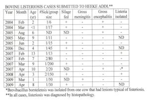
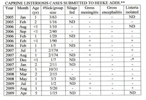
submitting herds. Available submission forms and case records for the Heeke
lab submissions (17 caprine, 12 bovine) were pulled and tabulated (see tables
at end of article). Of the 17 caprine cases, four (24%) involved outbreaks of
three or more animals in the flock,, five (29%) involved two animals, and eight
(47%) involved only one animal at the time of submission. In contrast, of the
12 bovine cases, none (0%) involved three or more animals, three (25%) involved
two animals, and nine (75%) involved only one animal at the time of
submission. Thus, goat herds appear more likely to suffer listeriosis
outbreaks that involve multiple animals.
Bovine listeriosis is often associated with the feeding of silage, presumably
spoiled silage. As in the case reported here, almost all cases of caprine
listeriosis that we have seen do not involve the feeding of silage. Dietary
information was provided for eight of the Heeke lab goat submissions and, in
all eight cases, silage was not a part of the diet (0% feeding silage). In
contrast, dietary information provided for 10 of the bovine cases suggested
that seven (70%) of those cases were associated with silage feeding. The source
of infection in the goat cases is undetermined, but is presumed to be spoiled
hay or feed that has been contaminated with dirt or feces, as the natural
reservoir for Listeria monocytogenes appears to be soil and mammalian GI
tracts. One previous study found that listeriosis was more prevalent in goats
that browsed heavily, as compared to goats consuming hay or pasture (Johnson,
1996). Our data provided no information about browse patterns. Another study
has suggested that venereal transmission may occur in goats (Wiedmann).
Merningitis was grossly visible in this case and, in my experience, is much
more likely to be present in cases of caprine listeriosis. In contrast,
meningitis is seldom noticeable in cases of bovine listeriosis. Gross
meningitis was reported in six (35%) of the caprine cases, but it was reported
in only one (8%) of the bovine cases. Although meningitis was more frequently
seen in goats, it is still not a reliable diagnostic finding. Interestingly,
grossly visible encephalitis (focal hemorrhage and necrosis of the brain stem)
was more frequently reported in the bovine cases. Four (31%) of the bovine
cases reported grossly visible encephalitis, whereas none (0%) of the goat
cases had grossly visible encephalitis.
It has been my impression that Listeria monocytogenes is more easily
isolated from the brains of affected goats, and a previous study suggests this
may be true (Johnson, 1995). Our records show a similar tendency, though less
dramatically. Listerial cultures were attempted in 14 of the goat cases and 10
bovine cases, and it was successfully isolated from four (29%) of the affected
goat brains, and from two (20%) of the affected bovine brains. In a fifth goat
case, we isolated an unidentified Corynebacterium-like species. Although this
isolate differed biochemically from the usual strains of Listeria
monocytogenes, positive immunohistochemistry results suggested that it was
antigenically related to Listeria. In one bovine case, we failed to
isolate Listeria, but Brevibacillus borstelensis was isolated
from the brain. The histologic lesion in this case was typical of listeriosis,
but it is possible that Brevibacillus was the actual cause of the
lesion. In both species, bacterial culture continues to be a relatively
unreliable diagnostic procedure for encephalitis listeriosis. The poor rate of
isolation may be due to prior treatment with antibiotics, as most of these
animals had reportedly been treated prior to death.
In our laboratory, most cases of listeriosis are diagnosed by histopathology
and, of the traditional diagnostic procedures, histopathology continues to be
the most reliable method of diagnosis for encephalitic listeriosis. Formalin
fixed sections of brain stem are the preferred sample to submit. (Please
remember that Listeria lesions occur only in the brain stem, and submission of
cerebral cortex may provide false negative results). Several newer and more
sensitive diagnostic procedures are also available at ADDL. As mentioned
above, we can diagnosis listeriosis by immunohistochemistry (IHC) and the
preferred sample again is a formalin-fixed section of brain stem. A previous
study found that IHC was much more sensitive than bacterial culture (Johnson,
1995). In addition, our molecular diagnostics lab can now diagnose listeriosis
by PCR analysis. For this, the preferred sample would be a segment of fresh
(unfixed) brain stem.
Our data provided no information about treatment of listeriosis, but treatment
is often unrewarding due to the difficulty of getting therapeutic levels to the
brain. Nevertheless, one previous study reported that, of nine animals treated
with a combination of gentamicin and ampicillin, six survived (Braun).
|
|
Photos provided by Dr. Jose
Ramos-Vara |
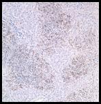
Detail of
immunohistochemical staining. There are numerous labeled bacteria within
hepatocytes. Immunoperoxidase- DAB for Listeria sp. |
Listeriosis.
Liver. Dark shading (brown) indicates the presence of Listeria antigen
within necrotic foci. Immunoperoxidase-DAB for Listeria sp. |
|
|
Corynebacterium
sp. (biochemically different from Listeria) was isolated from one brain,
but the brain stained positive for Listeria by immunohistochemistry.
** In all cases, listeriosis was
diagnosed by histopathology.
|
-by
Dr. Duane Murphy, ADDL PathologistHeeke ADDL
References