Diagnosis: Rhodococcus
equi in a horse
History: A 4-month-old
Tennessee walking horse colt was submitted to the Indiana Animal Disease
Diagnostic Lab at Purdue University for necropsy. The submitter reported an
approximately 1-1/2 month history of pneumonia and osteolytic lesions in the
right distal cannon bone and fetlock joint. The foal was treated with
antibiotics and had two arthroscopic surgeries on the affected joint, but did
not show significant signs of improvement and was subsequently euthanized.
Gross findings: The right
fetlock joint contained moderate amounts of cloudy tan to white fluid, with
numerous strands of fibrin. The synovial membranes were thickened and covered
by tan to yellow exudates that extended to and infiltrated into the deep
digital flexor and common digital extensor tendons as well as the interosseous
ligament. The distal physis of the cannon bone was lytic, had roughed
irregular surfaces, and was coated by tan to pink, thick, creamy material.
The lungs were diffusely mottled dark red to pink, wet, heavy, and oozed red
froth on cut section. Numerous (10-15) firm, tan to red, raised, round to
irregularly shaped nodules ranging in size from 2X2X1.5 - 8X8X6 cm were
scattered throughout all lung fields. The nodules contained abundant thick ,
tan to yellow, creamy exudate. The tracheobronchial lymph nodes ranged in
size from 3-7 cm in diameter and were tan to red, slightly firm and, when
incised, contained copious amounts of creamy tan material.
Histologic findings: The
distal physis of the cannon bone was segmentally separated from the epiphysis.
At the interface with the epiphysis, the physis was necrotic with a fibrillated
surface; there were decreased numbers of cells with few small clusters of
degenerate chondrocytes, and it was segmentally infiltrated by numerous
neutrophils and fewer macrophages. Marrow spaces of the metaphysis and
epiphysis were infiltrated largely by neutrophils with fewer macrophages and a
loose meshwork of fibrous connective tissue. Macrophages had abundant foamy
eosinophilic cytoplasm that was stippled with basophilic rod shaped bacteria.
Bony trabeculae had a scalloped surface, were multifocally fragmented, and
segmentally lacked a peripheral rim of osteoblasts. There were increased
numbers of osteoclasts along the peripheral trabeculae. Inflammatory cells
extended through the periosteum and into the adjacent soft tissues forming
pyogranulomas.
Pulmonary airways and adjacent alveolar spaces were multifocally effaced by
large aggregates of neutrophils, macrophages, and multinucleated giant cells.
The mononuclear cells contained variable numbers of round, eosinophilic, intracytoplasmic
bacteria (bacilli). Surrounding and coursing through the inflammatory
aggregates were thick mats of mature fibrous connective tissue. An area of
coagulative necrosis was focally circumscribed by clusters of neutrophils and
fibrous tissue, consistent with an abscess. Within the less severely affected
segments of lungs, the alveolar spaces contained numerous macrophages and fewer
neutrophils. Alveolar septa were segmentally lined by type II pneumocytes.
|
|
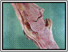
Cranial ventral aspect
of left lung lobe : pulmonary abscess |
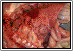
Right fetlock joint:
suppurative tenosynovitis and osteomyelitis |
Ancillary findings: A swab from the fetlock joint and sections of lung,
spleen, lymph node, and small intestine were submitted for aerobic bacterial
culture. Rhodococcus equi was isolated from the lungs, fetlock joint,
spleen, and lymph node. A goodpasture gram stain was applied to sections of
cannon bone and lung and revealed numerous, intracytoplasmic, gram positive
bacilli within macrophages.
Discussion: Characteristic
gross and microscopic lesions, coupled with positive bacterial cultures and
identification of intracytoplasmic, gram positive bacilli, were consistent with Rhodococcus equi infection in this foal. R. equi is a common
pathogen in foals that is predominantly seen in animals less than six months of
age. The primary target organs are the lungs, intestines, and associated lymph
nodes with resultant pyogranulomatous broncho-pneumonia, ulcerative
typhlocolitis, and pyogranu-lomatous lymphadenitis, respectively. Once the
infection is established, hematogenous dissemination may occur to the liver,
spleen, bone/joint, and skin. Additionally, R. equi has been documented
to cause abscesses in swine, sheep, cats, cattle, llamas.
R. equi is a gram positive
facultative intracellular bacteria that is found in soil. Most infections are
acquired through inhalation or ingestion of soil-borne organisms and virulence
is attributed to at least two predominant mechanisms. Replication occurs
within the cytoplasm of macrophages and is associated, at least in part, to the
expression of the plasmid-encoded, virulence-associated protein, VapA. Additionally,
virulence is achieved by preventing lysis through inhibiting the conversion of
phagosomes to phagolyosomes.
Antemortem diagnosis is based on clinical signs, evidence of pulmonary abscess
on thoracic radiographs, and aerobic bacterial cultures of transtracheal wash
fluid, abdominal fluid, or synovial fluid.
-by
Dr. Chad Frank, ADDL Graduate Student
References:
-
Caswell JL, Williams KJ: 2007.
Respiratory system. IN: Jubb, Kennedy and Palmer's Pathology of Domestic
Animals. 5th ed., Vol. 2. Ed. Maxie MG. Saunders-Elsevier,
Philadelphia, PA. pp. 630-632.
-
Hondalus MK: 1997.
Pathogenesis and Virulence of Rhodococcus equi. Veterinary Microbiology
56: 257-268.
-
Smith BP: 2002. Large Animal
Internal Medicine. 3rd ed. Mosby, St. Louis MO.
|