By Dr. Jose Ramos-Vara, ADDL Pathologist, Head of Histology
Immunohistochemistry (IHC) uses immunologic and histologic techniques to detect antigens in tissues. The antigen is recognized by a specific antibody that is added to the section. The immunologic reaction is visualized under the microscope by adding an enzyme, a substrate to the enzyme and a chromogen, producing a colored reaction. IHC is a very sensitive and specific technique. Advantages of IHC in diagnostics include: 1) Retrospective and prospective studies can be done on a variety of samples, 2) Antigen detection can be correlated with morphologic changes, 3) Stained slides can be stored for a long time, 4) Routine fixation and processing of samples is acceptable, 5) Tissues in paraffin blocks can be stored for years and still be suitable for immunohistochemistry.
Uses of IHC in veterinary diagnostics
Neoplastic and infectious diseases are the main focus of IHC in veterinary medicine. The Purdue ADDL IHC service offers a variety of tests for both infectious and neoplastic diseases of a variety of animal species. Please contact the ADDL for current tests available and fees or check the tests offered online (www.addl.purdue.edu/SampleSubmission/Immuno.aspx). Following are several examples in which IHC has practical application.
- Diagnosis of neoplasia. Often, the tissue origin of a tumor cannot be determined with routine histology. Using specific antibodies for different tissues or cells (e.g., cytokeratin for epithelium, vimentin for mesenchymal cells, lymphoid markers, etc.), the origin of many tumors can be determined with IHC. Immunohistochemistry is commonly requested for lymphoma (B vs T cells), histiocytic proliferations (e.g., histiocytic sarcoma), endocrine neoplasms, and undifferentiated neoplasms
- Detection of micrometastases. Early metastasis can be difficult to detect using conventional histology. IHC highlights the presence of single or small groups of neoplastic cells in metastatic sites. Early detection of micrometastases increases the chances of survival with surgical removal of affected nodes or by modification of the treatment protocol.
- Prognostic markers. Some proteins are expressed in neoplastic, but not in normal mature cells (e.g. embryonal proteins), expressed in neoplastic cells in larger amounts than in normal cells (e.g. cycle-related proteins), or structurally modified in neoplastic cells (mutant p53 protein). These changes may have prognostic significance in certain tumor types. For instance, the immunohistochemical detection of KIT protein in mast cell tumors of dogs has prognostic significance.
- Diagnosis of infectious diseases. Detection of antigens of an infectious agent with IHC has etiological significance. The advantage of IHC over microbiologic techniques is that antigen detection can be correlated with histopathologic changes and thus can confirm the significance of a particular bacterial or viral isolate obtained by other methods. Importantly, IHC does not require fresh or frozen tissues because it uses the same procedure as routine histology (formalin-fixed, paraffin-embedded tissues).
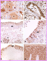
Fig. 1. Lymph node, pig. Porcine circovirus 2 infection. The antigen (brown color) is in the cytoplasm of syncytial (S) cells and histiocytes.
Fig. 2. Lung, equine fetus. Equine herpesvirus 1 infection. Intranuclear and intracytoplasmic labeling (brown color) of numerous cells of the bronchiolar (B) epithelium and alveolar (A) cells and macrophages.
Fig. 3 Nasal mucosa, dog. Canine distemper virus (CDV) infection. Numerous mucosal epithelial cells (ME) and the epithelium of the nasal glands (G) have CDV antigen (brown stain)
Fig. 4. Nasal mucosa, dog. Canine distemper virus (CDV) infection. Detail of CDV antigen in mucosal epithelial cells (ME).
Fig. 5. Lip, dog. Mycosis fungoides (epitheliotropic T-cell lymphoma). Neoplastic lymphocytes in the mucosal epithelium contain abundant CD3 antigen. CD3 is the marker for T lymphocytes.
Fig. 6. Lip, dog. Mycosis fungoides (epitheliotropic T-cell lymphoma).. Detail of the mucosal epithelium infiltrated by neoplastic CD3-positive lymphocytes.
How to submit samples for immunohistochemical testing
We test samples that have been fixed in formalin, so you do not have to do anything special. Just submit the sample as you would for routine histopathology. Please, do hot hold fixed samples in your office longer than 2 days, as prolonged fixation may destroy antigens. As soon as you place your sample in formalin, send it to ADDL. You are welcome to request specific tests that are available at ADDL. If not available at Purdue ADDL, we will try to find another laboratory performing that test. Alternatively, the diagnostician will suggest which test(s) are more appropriate after examining the HE sections on a given case. Currently, IHC tests are not included in the regular histopathology fee (IHC tests will accrue an additional cost), so we will contact you before performing IHC.
Interpretation of results
Immunohistochemistry facilitates diagnosis of infections and determination of the histogenesis and prognosis of neoplasms. Immunohistochemical results should be interpreted by the diagnostician provided that he/she has all the information pertaining to the case. For IHC, the diagnostician will send you a report indicating Positive (detected) or Negative (not detected) for a given marker (antigen) and, if pertinent, the percentage of positive cells and/or the antigen localization. Whether this result is significant must be interpreted in the context of the case, as is true for other diagnostic techniques. A careful assessment of the clinical history, lesions and all test results should be made before formulating a definitive diagnosis. Conversely, a negative result by immunohistochemistry does not eliminate the possibility of presence of a particular infectious agent/protein or its potential significance to the case. Sometimes infectious agents in subacute or chronic infections are in too low concentration to be detected by IHC. Due to mutations or other mechanisms, neoplastic cells may modify (upregulate/downregulate) the expression of proteins resulting in unexpected results.
In summary, immunohistochemistry is a valuable technique for the diagnosis of infectious and neoplastic diseases of animals. It is sensitive, specific, economical and relatively easy to perform. Although not always considered the “gold standard”, it can be as specific as bacterial and virus isolation, provided adequate controls are used.
References:
- Ramos-Vara JA: 2005. Technical aspects of immunohistochemistry. Veterinary Pathology 42:409-430.
- Ramos-Vara JA et al: 2008. Suggested guidelines for immunohistochemical techniques in veterinary diagnostic laboratories. Journal of Veterinary Diagnostic Investigation 20:393-413.