FINAL DIAGNOSIS: Rhododoccus equi in a goat
History: An 11-month old female Boer goat weighing 40 kg was submitted alive to the Purdue Animal Disease Diagnostic Laboratory for necropsy. The submitter reported an approximately two month history of anorexia, incoordination, and stilted gait. Other animals on the farm were not reportedly affected. At presentation, the animal was laterally recumbent, but could stand with assistance. If left alone, she would fall back into lateral recumbency. Sensation was present in all four limbs. There was no apparent head tilt or nystagmus.
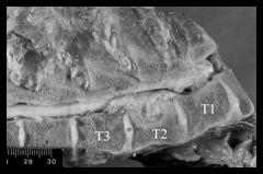
Gross examination: A tan to yellow, friable mass replaced approximately 50% of the dorsal aspect of the second thoracic vertebral body. The mass protruded into the spinal canal and compressed approximately three centimeters of the overlying spinal cord. The dura in this area was thin and red. Additional firm, white to tan, well-circumscribed nodules were present in the lung and liver. These nodules ranged in size from 2 mm to 12 mm in diameter and were lamellar on cut section. The left submandibular lymph node was enlarged. On section, the node architecture was replaced by friable, tan exudate.
Histopathologic examination: The vertebral body mass was a granuloma, composed of epithelioid macrophages and multinucleate giant cells that frequently contained large numbers of intracellular bacteria. The granuloma was surrounded by a thin layer of fibrous connective tissue. The adjacent bony trabeculae of the vertebral body were lytic and there was a concurrent suppurative osteomyelitis. The mass protruded into the spinal canal and extended slightly over the intervertebral disc. The dorsal aspect of the intervertebral disc and the physis of the vertebral body exhibited signs of degeneration. The overlying thoracic spinal cord was compressed. Many axons were swollen and surrounded by edematous myelin sheaths. Occasionally, myelin sheaths lacked axons and were infiltrated by macrophages. These axonal lesions were bilaterally symmetrical, limited to the area overlying the mass, and were most severe in the ventral funiculi. The submandibular lymph node, liver nodules, and lung nodules had similar morphology characterized by a necrotic center with multifocal dystrophic mineralization. The central necrotic mass was surrounded by epithelioid macrophages and multinucleated giant cells that contained numerous Gram’s- positive bacteria within cytoplasmic vacuoles. The nodules were encapsulated by a layer of fibrous connective tissue infiltrated with moderate numbers of neutrophils. The surrounding normal parenchyma of the organs was compressed.
Bacteriology: An abscess from the vertebral mass, along with samples of the liver, lung, and lymph node nodules, were submitted for aerobic bacterial culture. Rhodococcus equi was isolated from the vertebrae, liver and lung. Corynebacterium pseudotuberculosis was isolated from the submandibular lymph node.
Comment: The most common cause of disseminated granulomas or abscesses in sheep and goats is Corynebacterium pseudotuberculosis. When the disease is isolated to the lymph nodes, it is known as caseous lymphadenitis. While C. pseudotuberculosis was isolated from the lymph node, Rhodococcus equi was identified as the cause of the other granulomas. Rhodococcus equi was identified as the cause of the other granulomas. Rhodococcus equi is a common pathogen in foals where it can cause pneumonia, enteritis, abscesses, and osteomyelitis. Rhodococcus equi has also been documented to cause abscessed lymph nodes in swine, sheep, cats, cattle, and llamas. There have even been recent reports of Rhodococcus equi infecting immuno-suppressed human patients with HIV/AIDS. In goats, R. equi is reported to cause disseminated abscesses or granulomas, pneumonia, pleuritis, osteomyelitis, and lymphadenitis.
R. equi is a pleomorphic, Gram’s-positive, obligate intracellular bacterium most commonly residing in the soil where there is abundant avian or herbifore feces. The exact pathogenesis of R. equi infection in species other than foals is not completely understood. It is believed that infection may occur through ingestion or inhalation of the bacterium. Lesions in both the liver and lung of this animal would support both of these routes of entry. Once established, the organism may disseminate hematogenously throughout the body. Affected animals are largely asymptomatic, as is seen with caseous lymphadenitis, until the abscesses or granulomas compress and/or destroy vital tissues (i.e. spinal cord)
Based solely on gross and microscopic morphology, it is quite difficult to differentiate between abscesses caused by R. equi or Corynebacterium pseudotuberculosis. Even with a Gram’s stain, the morphology of the intracellular bacteria are similar. Bacterial culture is the most reliable method for differentiation. The high incidence of C. pseudotuberculosis and its characteristic gross appearance often discourage bacterial culture of these lesions when observed in the field. Because of this, the true prevalence of R. equi may be under-reported. Clinically, differentiation between the two organisms may be of benefit as the transmission and source of the organisms likely vary.
-by Dr. Robert Johnson, ADDL Graduate Student
|
A granuloma bulges from the second thoracic vertebra (T2) into the spinal canal where it compresses the spinal cord |
References:
-
Davis et al: 1999. Disseminated Rhodo-coccus equi infection in two goats. Veterinary Pathology 36(4): 336-339.
-
Kabonga et al: 2005. Caprine vertebral osteomyelitis caused by Rhodococcus equi. Journal of the South African Veterinary Association. 76(3): 163-164.
|